
Comprehensive Notes on Light and Electron Microscopes
Exploring the Tools that Illuminate the Microscopic World
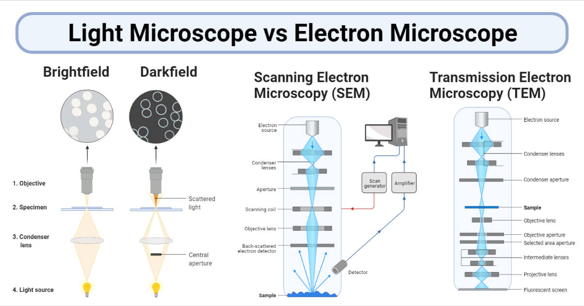
Key Takeaways
- Resolution and Magnification: Electron microscopes offer significantly higher resolution and magnification than light microscopes, enabling the visualization of structures at the nanometer scale.
- Operational Environments: Light microscopes operate in ambient conditions and can observe live specimens, whereas electron microscopes require vacuum environments and are limited to non-living samples.
- Applications and Accessibility: Light microscopes are widely accessible and used in educational and basic research settings, while electron microscopes are specialized tools essential for advanced scientific research and industrial applications.
Introduction
Microscopes are indispensable tools in the fields of biology, medicine, materials science, and numerous other disciplines. They enable scientists and researchers to observe structures that are invisible to the naked eye, fostering advancements in understanding and innovation. Two primary types of microscopes dominate scientific research: light microscopes and electron microscopes. Each operates on distinct principles, possesses unique components, and serves different applications based on their capabilities.
Light Microscopes
Definition and Principle
A light microscope, also known as an optical microscope, utilizes visible light and a series of lenses to magnify small objects. It operates by directing a beam of light through a specimen, which is then magnified by objective lenses and observed through an eyepiece. The fundamental principle relies on the bending of light rays to enlarge the image of the specimen, allowing for the visualization of cells, microorganisms, and other transparent or semi-transparent structures.
Key Components
1. Illumination System
The illumination system is critical for providing adequate lighting to view the specimen. It typically includes a light source such as a halogen or tungsten bulb, a condenser lens to focus light onto the specimen, and an adjustable diaphragm or iris to control light intensity and contrast.
2. Magnification System
The magnification system consists of multiple lenses that work together to enlarge the image of the specimen. This includes objective lenses with varying magnifications (commonly 4x, 10x, 20x, and 100x) and an ocular lens (eyepiece) usually with 10x magnification. The total magnification is determined by multiplying the magnification powers of the objective and ocular lenses.
3. Supporting System
The supporting system includes the stage, where the specimen is placed, and focus knobs (both coarse and fine) that adjust the distance between lenses and the specimen to achieve a sharp image.
Types of Light Microscopes
Light microscopes come in various types, each tailored for specific applications:
- Compound Microscopes: Utilize multiple lenses for higher magnification, ideal for viewing thin specimens like cells and bacteria.
- Stereomicroscopes: Provide lower magnification with a three-dimensional view, suitable for larger, opaque specimens.
- Fluorescence Microscopes: Employ specific wavelengths to excite fluorescent dyes in specimens, revealing detailed structures.
- Phase-Contrast Microscopes: Enhance contrast in transparent specimens without the need for staining.
- Polarized Light Microscopes: Utilize polarized light to study anisotropic materials such as crystals.
Advantages
- Ability to observe live and dynamic specimens, facilitating the study of biological processes in real-time.
- Relatively affordable and accessible, making them common in educational and basic research laboratories.
- Simple and straightforward sample preparation with established techniques for staining and labeling.
Disadvantages
- Limited resolution (~200 nanometers) due to the wavelength of visible light, restricting the observation of very small structures.
- Lower magnification capabilities compared to electron microscopes, limiting the study of nanoscale features.
- Some specimens may lack contrast without specialized staining techniques, making visualization challenging.
Applications
- Biological and medical research for examining cell structures, tissues, and microorganisms.
- Educational settings for teaching basic microscopy techniques and fundamental biological concepts.
- Material science for optical examination of surfaces and studying the properties of various materials.
Electron Microscopes
Definition and Principle
Electron microscopes utilize beams of accelerated electrons instead of visible light to form images of specimens. Because electrons have much shorter wavelengths than photons, electron microscopes achieve significantly higher resolution and magnification, enabling the visualization of structures at the nanometer and even atomic scale. The primary principle involves the transmission or scanning of electrons through or over the specimen, with electromagnetic lenses focusing the electron beam to produce detailed images.
Key Components
1. Electron Source
The electron source, often an electron gun with a heated tungsten filament, generates and accelerates the electron beam. Electrons are emitted through thermionic emission and then accelerated towards the specimen using electric fields.
2. Imaging System
The imaging system comprises electromagnetic lenses that manipulate the electron beam, focusing it onto the specimen. In Transmission Electron Microscopes (TEM), electrons pass directly through the specimen to form an image on a photographic plate or detector. In Scanning Electron Microscopes (SEM), the electron beam scans the specimen's surface, and detectors capture secondary electrons to create detailed surface topographies.
3. Vacuum Chamber
Electron microscopes operate within a high vacuum environment to prevent electrons from scattering off air molecules, which would degrade image quality and limit resolution.
Types of Electron Microscopes
- Transmission Electron Microscope (TEM): Transmits electrons through ultra-thin specimens to reveal internal structures with high resolution, allowing magnifications up to 2 million times.
- Scanning Electron Microscope (SEM): Scans a focused electron beam over the specimen's surface, producing three-dimensional-like images with detailed surface topology.
Advantages
- Extremely high resolution, capable of resolving structures down to the nanometer and atomic levels.
- Greater depth of field in SEM provides detailed three-dimensional views of specimen surfaces.
- Ability to analyze composition through techniques like Energy-Dispersive X-ray Spectroscopy (EDS), enabling elemental analysis of specimens.
Disadvantages
- Specimens must be placed in a vacuum, making it impossible to observe living organisms.
- Extensive and complex sample preparation is required, including dehydration, fixation, and coating with conductive materials.
- Electron microscopes are expensive to purchase and maintain, necessitating specialized facilities and trained personnel.
- Images produced are typically grayscale, lacking the natural color seen in light microscopy.
Applications
-
Materials science for studying crystallography, nanotechnology, and metallurgy properties.
-
Biological research for examining cellular ultrastructure, virus morphology, and detailed tissue organization.
-
Semiconductor industry for the inspection and failure analysis of microelectronic components.
-
Forensic science for detailed analysis of evidence materials at the microscopic level.
Comparison Between Light and Electron Microscopes
| Feature | Light Microscope | Electron Microscope |
|---|---|---|
| Illumination Source | Visible light (400-700 nm) | Electron beam (~1 nm wavelength) |
| Lens Type | Glass lenses | Electromagnetic lenses |
| Operating Environment | Ambient conditions, no vacuum required | High vacuum environment |
| Specimen Preparation | Minimal, can observe live specimens, staining possible | Complex, requires fixation, dehydration, and conductive coating |
| Magnification | Up to 1,500x | Up to 1,000,000x |
| Resolution | ~200 nanometers | Up to 0.001 micrometers |
| Image Viewing | Directly through eyepiece, color images | On photographic plates or screens, typically grayscale |
| Advantages | Ease of use, affordability, ability to view live specimens | High resolution and magnification, detailed surface and internal imaging |
| Disadvantages | Limited resolution and magnification, lower depth of field | Expensive, cannot view live specimens, complex operation |
| Applications | Biological studies, education, basic material examination | Advanced scientific research, materials science, semiconductor inspection |
Conclusion
Light and electron microscopes are fundamental instruments in scientific exploration, each offering unique advantages and applications based on their intrinsic capabilities. Light microscopes serve as accessible and versatile tools for educational purposes and basic research, enabling the observation of live and dynamic biological specimens with minimal preparation. In contrast, electron microscopes provide unparalleled resolution and magnification, essential for advanced research in fields requiring detailed imaging at the nanoscale. However, the complexity, cost, and operational requirements of electron microscopes limit their accessibility compared to their light counterparts. Understanding the strengths and limitations of each microscope type allows researchers to select the most appropriate tool for their specific investigative needs, thereby advancing scientific knowledge and technological innovation.
References
Last updated February 12, 2025