
Dermatology Terminologies: A Comprehensive Glossary
Explore essential dermatology vocabulary excluding disorders, tests, and treatments
Highlights
- Foundational Skin Anatomy: Understand the epidermis, dermis, hypodermis, adnexae, and associated structures.
- Lesion Descriptions & Morphology: Explore detailed descriptors such as papules, plaques, macules, and scales.
- Descriptive Terminologies: Familiarize with terms related to distribution, configuration, and visual/tactile descriptions like erythema, hyperpigmentation, and lichenification.
Introduction to Dermatology Terminologies
In dermatology, precise vocabulary is essential for effective communication among healthcare professionals. This glossary focuses on terminology related to skin anatomy, descriptive evaluations, and terms used during clinical assessments. These terms form the cornerstone of dermatological diagnostics and greatly assist in the clear documentation of skin features.
Fundamental Skin Anatomy
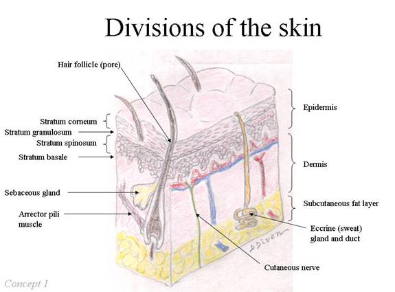
The skin is the largest organ in the human body and can be divided into several layers and associated structures. Each component plays a critical role in protecting the body, regulating temperature, and serving as a sensory interface with the environment. The primary layers include:
Epidermis
The epidermis is the outermost layer of the skin. It consists mainly of keratinocytes, which are cells that produce keratin, a protein that helps form a barrier against environmental damage, pathogens, and water loss. Within the epidermis, specialized cells such as melanocytes synthesize melanin, the pigment responsible for skin color and protection against ultraviolet radiation.
Dermis
Situated beneath the epidermis, the dermis is a thicker layer composed of connective tissue, blood vessels, nerve endings, hair follicles, and sweat glands. This layer provides structural support and elasticity to the skin. It is subdivided into two regions: the papillary dermis (upper) and the reticular dermis (lower).
Hypodermis (Subcutaneous Tissue)
The hypodermis is not technically part of the skin but lies immediately beneath the dermis. It primarily consists of adipose (fat) tissue that provides insulation, energy storage, and cushioning for the underlying muscles and organs.
Adnexae
The term "adnexae" refers to accessory skin structures such as hair follicles, sebaceous glands, and sweat glands. These structures contribute to various skin functions, including thermoregulation, lubrication, and protection against infections.
Descriptive Terminology in Dermatological Assessment
When dermatologists examine the skin, they utilize a pool of descriptive terms that detail the morphology, distribution, configuration, and surface characteristics of skin lesions. This lexicon aids in forming a systematic assessment of the clinical presentation without implying a specific disease.
Lesion Terminology
A lesion is defined as any area of the skin that differs in appearance from the surrounding tissue. Lesions can vary significantly in their characteristics:
Macro Descriptors: Size and Shape
Lesions are described in terms of their size, shape, and nature:
- Macule: A flat, distinct, discolored area of skin less than 1 cm in diameter. Since it is flat, it is only identifiable by a change in color.
- Patch: A flat discolored area larger than a macule; typically greater than 1 cm.
- Papule: A small, raised, solid bump on the skin, often less than 1 cm in diameter.
- Plaque: A raised, flat-topped lesion typically larger than a papule, often appearing as a plateau-like elevation.
- Scale: Flaky, crusted, or scaly surface that results from the shedding of the outer layer of the epidermis.
Textural and Color Descriptors
In addition to size, dermatologists often evaluate visual and tactile characteristics:
- Erythema: Redness of the skin due to enhanced blood perfusion; this is commonly observed during inflammatory reactions.
- Hyperpigmentation: Darkening of the skin beyond its normal color, caused by an excess of melanin production.
- Hypopigmentation: A lighter area of skin resulting from reduced melanin.
- Leukoderma: Areas of significant skin lightening, sometimes used interchangeably with achromia.
- Violaceous: Describes a purple hue observed in skin, which may be due to an accumulation of blood or certain vascular changes.
Spatial Distribution and Arrangement
A critical aspect of dermatological descriptions is the spatial arrangement of lesions. These descriptors help to visualize how skin changes are distributed:
Distribution
Distribution refers to the pattern by which lesions appear on the body—whether they are localized, generalized, symmetric, or follow specific patterns such as dermatomal (involving the area supplied by a single spinal nerve) or linear.
Configuration
Configuration details the arrangement of lesions in relation to each other. Descriptors include:
- Annular: A circular or ring-like configuration of lesions.
- Linear: Lesions aligned in a straight or near-linear pattern, often indicative of external forces or rubbing.
- Clustered: Lesions that are grouped together in a localized area.
Additional Descriptive Terms
Aside from categorizing lesions by size and arrangement, further terminology enriches descriptions regarding surface features and subtle characteristics:
Surface Characteristics
Skin surface features offer vital clues about the physiological processes:
- Blanching: The temporary loss of color when pressure is applied to the skin, indicating capillary refill and blood flow status.
- Desquamation: The process of skin peeling or shedding the outer layer, often as a response to irritation or as part of a healing process.
- Lichenification: Thickened, rough skin with accentuated skin markings, usually resulting from chronic friction or rubbing.
- Crusting: Formation of a hardened layer on the skin surface due to dried exudates such as blood or pus.
- Erosion: A superficial loss or break in the epidermis resulting in an area where the skin is missing, though not affecting deeper layers.
Anatomical and Technical Terms
Several technical terms in dermatology pertain to the underlying structure and accessory components of the skin:
- Adnexal Structures: Refers to the hair, nails, and glandular components that are integral to skin functions.
- Sebum: An oily secretion from sebaceous glands that lubricates and waterproofs the skin.
- Stratum Corneum: The outermost layer of the epidermis, consisting of dead, keratinized cells which provide a barrier function against mechanical and chemical insults.
- Follicular: Pertaining to hair follicles and their structures.
- Cosmesis: While not strictly an anatomical term, this refers to aspects of appearance and physical beauty as preserved or enhanced through cosmetic techniques.
Comprehensive Glossary Table
The following table summarizes key dermatology terminologies, their definitions, and relevant application contexts in clinical descriptions.
| Term | Definition | Clinical Context |
|---|---|---|
| Adnexae | Accessory structures like hair follicles, sweat glands, and sebaceous glands. | Aid in describing the skin’s accessory components. |
| Epidermis | The outer layer of the skin, composed primarily of keratinocytes. | Describes the protective barrier and pigmentation processes. |
| Dermis | The underlying layer containing connective tissue, blood vessels, and nerve endings. | Focuses on the elastic and supportive properties of the skin. |
| Hypodermis | A fatty layer beneath the dermis that provides insulation and cushioning. | Important for understanding subcutaneous features and energy storage. |
| Macule | A small, flat discolored area of skin less than 1 cm in diameter. | Used to note color changes in the skin’s appearance. |
| Patch | A larger flat area, typically more than 1 cm, with altered coloration. | Helps differentiate superficial changes in skin pigment. |
| Papule | A small raised bump on the skin. | Commonly noted in inflammatory or reactive skin conditions. |
| Plaque | A raised, flat-topped lesion, often larger than papules. | Indicates sustained elevations on the skin’s surface. |
| Scale | Flaky or dry skin areas resulting from shedding of the epidermal layer. | Describes surface texture and potential desquamation. |
| Erythema | Redness of the skin caused by increased blood flow. | Used to annotate inflammatory or irritative processes. |
| Hyperpigmentation | Increased skin pigmentation resulting in darker areas. | Important for assessing melanin-related changes. |
| Hypopigmentation | Reduced pigmentation leading to lighter skin areas. | Noted in conditions with diminished melanin. |
| Leukoderma | Marked loss of skin pigment, often creating stark white patches. | Serves as an extreme form of hypopigmentation. |
| Violaceous | A purple hue indicating vascular changes or blood stasis. | Often used in contexts where vascular phenomena are present. |
| Annular | Ring-like or circular lesions. | Helps to conceptualize the pattern of skin changes. |
| Linear | Lesions arranged in a straight or nearly straight line. | May suggest an external cause such as trauma or scratching. |
| Blanching | Temporary loss of skin color when pressure is applied. | Used as an indicator of blood flow and capillary health. |
| Desquamation | Peeling or shedding of the outer skin layer. | Observed in healing processes or after irritation. |
| Lichenification | Thickened, rough skin often due to persistent friction. | Marks chronic skin irritation or rubbing. |
| Crusting | The formation of a dried layer of blood, pus, or other fluids on the skin. | Noted in response to exudative skin conditions. |
| Erosion | A superficial loss of the epidermis resulting in an open area. | Helps differentiate from deeper ulcerative processes. |
In-Depth Discussion of Key Concepts
Morphology and Clinical Descriptions
Morphology in dermatology is paramount as it relates to the description of the visual characteristics of skin lesions. Assessing the morphology entails a thorough examination of both the primary features (such as size, shape, color, and texture) and the secondary features (such as surface changes, erosions, or crusting). Clinicians utilize these descriptors in a systematic way to ensure precise communication whether assessing changes over time or discussing findings with colleagues.
For instance, a lesion may be described as a “linear papule with surrounding erythema,” directly conveying the lesion’s shape (linear), size and elevation (papule), and color aspect (erythema). Such detailed descriptions help in documenting the progression of skin conditions and in formulating differential diagnoses without implying any direct etiology.
Descriptive Terminologies in Practice
In clinical practice, descriptive terminologies enable practitioners to differentiate normal skin variations from pathological changes. Consider a scenario where multiple lesions are present; the clinician would document the following:
- A detailed account of the lesion's morphology – noting whether the lesions are macular (flat) or papular (raised).
- An evaluation of distribution patterns – determining if the lesions occur in a symmetric, unilateral, or dermatomal pattern.
- An analysis of configuration – for example, whether lesions are annular or arranged in a linear pattern which might suggest external irritation.
- Surface details such as the presence of scaling, crusting, or desquamation, which may indicate a reparative or infectious process.
These elements ensure that the information captured is comprehensive enough for both the immediate clinical context and for future correlational studies on skin disorders. This approach underpins the systematic nature of dermatological examinations and is essential in multi-disciplinary communications.
Utilization of Technical Terminologies
Dermatologists often rely on a technical vocabulary that includes anatomical terms as well as dynamic terms used during skin examinations. For instance, when evaluating a suspected inflammatory process, the practitioner might comment on the visible "erythema" and assess "blanching" during pressure tests. Similarly, subtle changes such as "hyperpigmentation" or "hypopigmentation" can indicate variations in melanin production without necessarily being predetermined as disease indicators.
The use of specific terms like "adnexae" underscores the importance of recognizing accessory structures and their roles. This detail is critical when planning any interventions that might involve cosmetic procedures or when discussing potential side effects of dermatological conditions.
Additional Notes on Dermatological Vocabulary
It is important to note that the vocabulary used by dermatologists is not only descriptive but also highly standardized. This standardization aids in research, teaching, and the global conversation surrounding skin conditions. Terms such as "follicular" and "cosmesis" emphasize not only the structural descriptions but also considerations regarding appearance and aesthetic outcomes.
Furthermore, many of these terms extend into other medical fields, showing interdisciplinary relevance. For example, "blanching" is used in both dermatology and vascular medicine to assess the health of capillaries. Similarly, the concept of "lichenoid" changes finds parallels in pathology where chronic rubbing leads to observable skin thickening.
The systematic assessment of skin conditions—focusing on aspects like lesion morphology, distribution, and surface changes—is foundational to dermatological practice. Whether communicating findings in academic research or clinical practice, these standardized terms allow for precise descriptions that facilitate meaningful comparisons across patient populations and over time.
References
- Dermatology Terms - American Osteopathic College of Dermatology
- Terminology in Dermatology - DermNet NZ
- Glossary for Dermatology Terms - ILDS
- Glossary of Dermatology Terms - UCI Health
- Dermatology Glossary of Terms - Soothems
Related Queries for Further Exploration
Last updated March 6, 2025