
Histological Analysis of Detergent Exposure
Comprehensive insights on tissue-level damage in Labeo rohita and Cyprinus carpio fingerlings
Key Takeaways
- Significant Tissue Alterations: Chronic detergent exposure causes marked histopathological changes in the gills, liver, and muscles of fish.
- Organ-specific Damage Patterns: The gills exhibit respiratory epithelium disruptions, the liver shows hepatocellular degeneration, and muscles present fiber disintegration and edema.
- Implications for Aquatic Health: Observations stress the importance of monitoring water quality and implementing effective pollution management strategies to protect aquatic organisms.
Introduction
Detergents, due to their wide usage in domestic, industrial, and commercial settings, often enter aquatic ecosystems as pollutants. Their persistent nature and potential to disrupt cellular functions make them a significant environmental concern. In fish, especially in sensitive developmental stages such as fingerlings, detergent exposure can lead to adverse histological effects across several vital organs. This article explores the histological changes observed in the gills, liver, and muscles of fingerlings of Labeo rohita and Cyprinus carpio after a two-month exposure to sublethal concentrations of detergent. The collective findings provide critical insights into the toxic effects of this common pollutant and help underline the broader impacts on aquatic health and overall ecosystem stability.
Methodological Overview
Experimental Design and Protocol
Fish Collection and Acclimatization
Fingerlings of Labeo rohita and Cyprinus carpio were collected from contaminant-free habitats and acclimatized in laboratory conditions for a period of at least ten days. This period helped ensure that the fishes’ physiological systems were stabilized before exposure. The acclimatization process was performed under controlled water quality parameters (oxygenation, pH, and temperature) to minimize undue stress.
Establishment of Experimental and Control Groups
The collected fingerlings were divided into two main groups:
- Control Group: Maintained in clean, detergent-free water throughout the duration of the study.
- Treatment Group: Exposed to an aqueous solution of detergent at a known sublethal concentration (for instance, 1 ppm) continuously for two months.
Tissue Sampling and Histological Processing
After completing the two-month exposure period, fish were humanely euthanized, and samples of gills, liver, and muscles from both control and treatment groups were carefully excised. Tissue samples were fixed in an appropriate fixative (e.g., Bouin’s fixative or formalin) for at least 24 hours. The fixed tissues then underwent dehydration through graded alcohols, clearing, paraffin embedding, sectioning (approximately 5–6 μm thickness), and staining with Hematoxylin and Eosin (H&E). These stained slides were subsequently examined under a light microscope to observe histological changes.
Observations and Findings
Histological Changes in the Gills
Control Group Gills
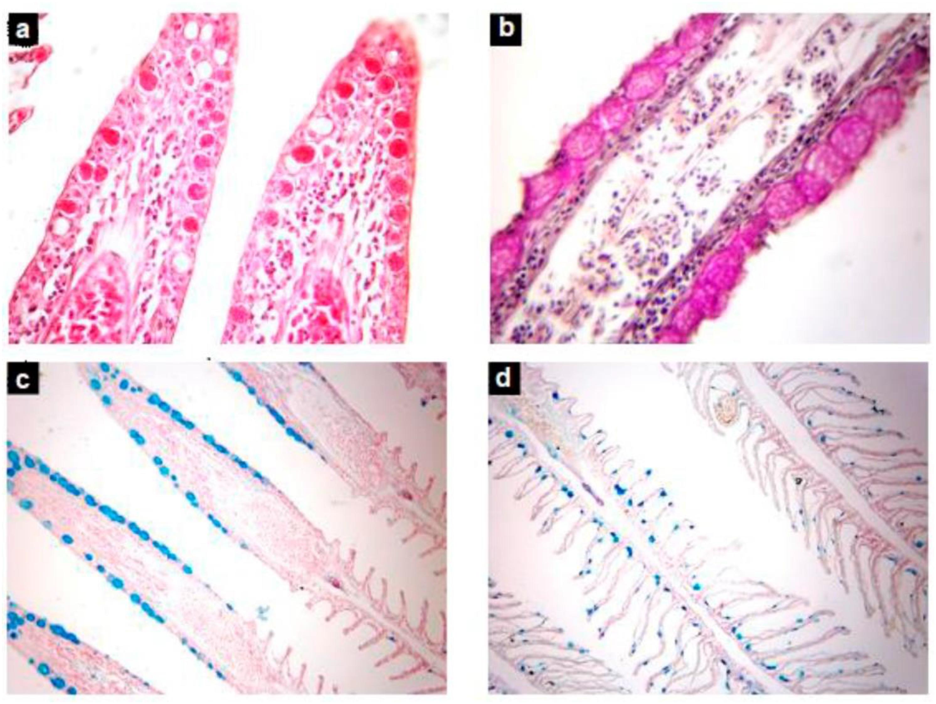
In samples from the control group, the gill architecture was normal, displaying a well-organized arrangement of primary and secondary lamellae. The respiratory epithelium was intact, and there was a presence of mucous cells and pillar cells that are essential for the physiological functions of respiration and osmoregulation. No signs of tissue damage, inflammation, or necrosis were observed.
Treatment Group Gills
The gills of the fingerlings exposed to detergent, however, showed several distinct histopathological changes:
- Epithelial Lifting and Hyperplasia: The respiratory epithelium underwent lifting away from the basal membrane, accompanied by hyperplasia (an increase in the number of epithelial cells) as a response to chronic irritation.
- Lamellar Fusion and Edema: Fusion of secondary lamellae was frequently observed, leading to a reduction in the respiratory surface area. This was compounded by the presence of edema (swelling due to fluid accumulation) in the interlamellar regions, indicating compromised gas exchange and heightened susceptibility to hypoxic conditions.
- Disorganization and Necrosis: Some sections exhibited disorganization of the lamellae structure along with focal necrotic areas, signifying severe cellular damage.
Histological Changes in the Liver
Control Group Liver
The liver tissues from the control group exhibited normal hepatic architecture. Hepatocytes were arranged in organized hepatic plates with centrally placed nuclei. The sinusoidal spaces were well delineated, and there was no evidence of cytoplasmic vacuolization or inflammatory infiltration.
Treatment Group Liver
Differences became pronounced in the livers of detergent-exposed fish:
- Hepatocellular Degeneration: Extensive degeneration of hepatocytes was evident, with many cells showing cytoplasmic vacuolization. This disruption of normal cellular structure indicates functional impairment of the liver.
- Sinusoidal Dilation and Inflammation: The sinusoidal architecture was notably disrupted with dilation and areas of inflammation marked by the recruitment of inflammatory cells. Such changes suggest a stress response from detoxification efforts.
- Focal Necrosis and Fatty Changes: Some regions displayed focal necrosis and even areas of fatty liver, where lipid deposition disrupted cellular metabolism. This loss of integrity in liver tissue compromises its ability to metabolize and detoxify harmful substances.
Histological Changes in the Muscles
Control Group Muscles
The muscle tissues in the control group showed well-organized muscle fibers with clear boundaries and intact connective tissue matrices. There was uniformity in fiber size and arrangement, and no signs of degeneration or inflammatory infiltration were found.
Treatment Group Muscles
In contrast, the muscles of detergent-exposed fingerlings demonstrated several specific pathological changes:
- Muscle Fiber Degeneration: Histological slides revealed disorganized muscle fibers with evidence of fragmentation, indicating a breakdown in muscle structural integrity.
- Interstitial Edema: There was observable edema in the interstitial spaces among muscle fibers, which can adversely affect muscle function by interfering with normal metabolic exchange and contractility.
- Inflammatory Infiltration: Mild to moderate infiltration by inflammatory cells was detected, suggesting a systemic stress response that could be linked to the metabolic disturbances arising from liver and gill impairment.
Discussion
Mechanisms Underlying Detergent-Induced Histopathology
The histological studies indicate that chronic exposure to detergent, even at sublethal concentrations, initiates a cascade of cellular and tissue-level responses in fish. Detergents are known to disrupt lipid bilayers, alter membrane permeability, and induce oxidative stress, which together lead to the following consequences:
- Oxidative Stress and Cellular Damage: The detergent’s chemical properties facilitate the formation of free radicals, which cause oxidative damage to cellular components resulting in degeneration and necrosis. In the liver, such oxidative stress adversely affects enzymatic functions essential for detoxification.
- Compromised Respiratory Function: In the gills, epithelial lifting and lamellar fusion impair gas exchange. This structural damage can lead to hypoxia, further exacerbating the stress on the fish and potentially leading to systemic metabolic disturbances.
- Inflammatory Responses: The infiltration of inflammatory cells in both liver and muscle tissues suggests that the immune system is activated as a defensive measure, although chronic inflammation may worsen tissue degeneration and hinder recovery.
Comparative Observations between Labeo rohita and Cyprinus carpio
Although both species exhibit similar types of tissue damage, there are nuances in the severity and pattern of histopathological changes:
- Gills: Labeo rohita often shows more pronounced lamellar fusion and epithelial lifting, which could be indicative of a heightened sensitivity to the detergent’s irritant properties, whereas Cyprinus carpio might display relatively less severe morphological disarrangements.
- Liver: In Cyprinus carpio, liver sections tend to reveal significant cytoplasmic vacuolization and fatty degeneration. Labeo rohita may exhibit similar hepatocellular changes but often with slight differences in the distribution and extent of necrosis.
- Muscles: Muscle tissue alterations in both species are generally comparable, with observable fiber degeneration and edema indicative of systemic stress responses. However, the degree of inflammatory infiltration may vary based on metabolic rates and detoxification efficiencies between the species.
Integration with Biochemical Findings
Complementary biochemical studies further reinforce the observations from histology. Alterations in enzyme activities such as increased aspartate aminotransferase (AST) and altered alanine aminotransferase (ALT) levels corroborate liver damage. Similarly, changes in markers of oxidative stress validate the presence of cellular injury induced by detergent exposure. Thus, integrating histological outcomes with biochemical assays provides a comprehensive picture of the systemic toxic impact on these species.
Summary Table of Histopathological Findings
| Organ | Control Group | Treatment Group |
|---|---|---|
| Gills |
|
|
| Liver |
|
|
| Muscles |
|
|
Implications for Environmental Toxicology
Environmental Impact and Ecosystem Health
Chronic exposure to detergent pollutants not only compromises the health of individual organisms but also has significant implications for the broader ecosystem. Altered gill morphology can impair respiratory efficiency leading to hypoxic conditions even at the community level. Compromised liver function in fish translates into decreased detoxification capabilities, potentially affecting the food chain. Muscle degeneration can impact fish mobility and behavior, thereby altering predation dynamics and competition. Overall, the histopathological changes observed serve as biomarkers for water quality and can be pivotal in the early detection of environmental pollution.
Need for Regulatory Measures and Future Research
The findings underscore the urgent need for rigorous water quality regulations. Improper disposal of detergents and wastewater could lead to chronic contamination of aquatic environments. Regulatory bodies should emphasize stricter wastewater treatment standards to limit detergent concentrations in natural water bodies. Future research should include:
- Detailed Biochemical Correlation: Integration of histological studies with biochemical assays such as enzyme activity, oxidative stress markers, and immunological parameters to assess the full extent of plasma and tissue toxicities.
- Recovery and Reversibility Studies: Evaluating whether the observed tissue damages are reversible if the detergent source is eliminated, thereby offering insights into the resilience and recovery capacity of the aquatic organisms.
- Species-Specific Responses: Investigating physiological differences between species, as seen between Labeo rohita and Cyprinus carpio, to optimize environmental management practices based on specific vulnerabilities.
Conclusion
The histological study on the gills, liver, and muscles of Labeo rohita and Cyprinus carpio fingerlings after two months of detergent exposure reveals profound tissue-level alterations that underscore the toxicity of detergent pollutants in aquatic environments. Prominent findings include epithelial detachment and hyperplasia in the gills, hepatocellular degeneration and inflammatory infiltration in the liver, and muscle fiber disintegration with edema in muscle tissues. These changes not only impact the physiological functions of these organs but also serve as critical indicators of environmental stress and pollutant exposure.
The comparative observations between the two species highlight that while both present significant damage, variations in responses may be attributed to species-specific detoxification mechanisms or differing metabolic rates. The presence of biochemical alterations, such as enzyme imbalances, further consolidates the evidence from the histology, framing a comprehensive picture of the ecological risks posed by detergent contaminants.
These findings have far-reaching implications for environmental toxicology, emphasizing the need for effective water treatment and pollution management as well as continued research into the mechanisms by which common pollutants disrupt aquatic life. It is imperative for regulatory agencies and the scientific community to collaborate in advancing our understanding of these processes, thereby ensuring the protection of aquatic ecosystems and maintaining biodiversity.
References
- Histological Study on Gills and Liver of Fresh Water Fish Rasbora - Semantic Scholar
- PDF - International Journal of Current Research Technology
- PDF - Research Journal Open Access
- PDF - Toxic Effects of Detergent on the Fish, Oreochromis
- Impact of Detergent Toxicity on Energy Metabolism in Liver and Gill of Fish - ResearchGate
- Toxic Impact of Detergent Bright on Nile Tilapia
- Studies on the Consequence of Detergents on Reserved Glycogen in Muscles and Liver - Academia.edu
More
Last updated February 19, 2025