
Comparative Histological Analysis of Rat and Human Stomach
Exploring the structural similarities and differences in gastric histology.
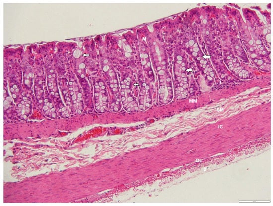
Highlights
- Structural Similarities: Both species share a basic four-layer organization including mucosa, submucosa, muscularis externa, and serosa.
- Species-Specific Differences: Unique features in the rat stomach such as the forestomach, limiting ridge, and differences in muscular structure significantly impact functional outcomes.
- Research Implications: Understanding these differences aids in translating findings from rat models to human gastric pathologies and treatments.
Introduction
The study of histology allows researchers to explore the microscopic structure of tissues in a detailed manner. In particular, comparing the histology of the rat stomach with that of humans has proven invaluable in biomedical research. Rats are widely used as experimental models due to the overall similarity of their gastrointestinal tract with that of humans. However, key differences exist that must be accounted for when extrapolating data.
In the context of translational research, as demonstrated by studies in cardiovascular disease, where findings from controlled trials such as the HOPE study have informed therapeutic approaches, the detailed characterization of the rat stomach paves the way for understanding human gastric pathologies. Similar to how ACE inhibitors were shown to have antiatherosclerotic properties and reduce myocardial infarction rates in patients with left ventricular dysfunction, studies in comparative gastric histology can help identify the pathophysiological underpinnings of gastrointestinal disorders. This reference aims to provide an in-depth comparative analysis of the histological features of rat and human stomachs, elucidating both the common morphological structures and the distinctive features that set these models apart.
Background and Rationale
The gastrointestinal tract’s functionality is critically dependent on its structural organization. Both rats and humans exhibit a four-layer organization of the stomach wall: the mucosa, submucosa, muscularis externa, and serosa. This structural complexity facilitates various functions such as mechanical digestion, secretion of digestive enzymes, and protection against the corrosive effects of gastric acid.
Historically, the anatomical and histological comparison between rats and humans emerged from the need to validate animal models in preclinical research. Understanding the detailed histological differences, such as the presence of a non-glandular forestomach in rats, is essential for interpreting results from experimental studies. Because features such as a prominent limiting ridge in rats and the unique distribution of cell types within the mucosal layer may influence how substances are processed in the stomach, researchers must consider these variations when designing studies and interpreting outcomes.
Methodology and Experimental Design
Sample Collection and Preparation
Tissue samples are typically harvested from both rat and human stomachs following ethical collection practices. For rats, standardized protocols ensure anesthesia and euthanasia are conducted in compliance with animal welfare guidelines. In human studies, samples may be obtained during surgical procedures or from autopsy specimens, with proper consent and ethical review.
Fixation and Staining
The collected tissues are immediately fixed in formalin to preserve cellular architecture. Subsequent processing includes embedding in paraffin, sectioning, and staining. Common stains such as hematoxylin and eosin (H&E) are used to highlight general tissue structure, whereas specialized stains (e.g., Periodic Acid-Schiff) can aid in the examination of mucopolysaccharides in mucus-secreting cells.
Microscopic Analysis
Histological slides are examined using light microscopy to assess the organization of layers, cellular composition, and the manifestation of specialized structures. Comparative analysis focuses on identifying common features such as the classification of cell types (parietal cells, chief cells, mucous cells) and differences such as the differential thickness and cellular arrangement between species.
Comparative Parameters
The analytical approach integrates several parameters:
- Tissue Architecture: Comparison of the general four-layer structure in both species.
- Cellular Composition: Detailed analysis of the distribution and density of parietal cells, chief cells, and mucus cells.
- Specialized Regions: Examination of the rat’s forestomach and limiting ridge versus the exclusively glandular organization in humans.
- Functional Correlations: Assessing how structural differences impact physiological outcomes such as acid secretion, motility, and the stomach’s capacity to launch protective responses.
Histological Findings: A Comparative Analysis
Overall Structural Organization
Both rat and human stomachs consist of four basic layers:
1. Mucosa: This innermost lining contains an epithelium that participates in secretory functions. In both species, the mucosal layer is subdivided into the surface epithelium, lamina propria, and muscularis mucosae.
2. Submucosa: A supportive tissue that contains blood vessels, nerves, and connective tissue, it aids nutrient exchange and provides structural support.
3. Muscularis Externa: Responsible for the contractile activity necessary for gastric mixing and propulsion, it is composed of three layers of muscle – longitudinal, circular, and oblique (in some regions).
4. Serosa: The outermost layer that provides a smooth surface, facilitating organ movement within the abdominal cavity.
Species-Specific Features
In rats, the stomach is divided into distinct regions. The anterior portion, known as the forestomach, is non-glandular and lined with stratified squamous epithelium. This region primarily serves as a storage area for ingested food, and its robust epithelium provides considerable resistance to mechanical stress. The forestomach is separated from the glandular region by a pronounced limiting ridge, which not only demarcates the two regions but also serves a functional role in preventing regurgitation by limiting retrograde movement of the contents.
In contrast, the human stomach is uniformly glandular from the cardia through to the pylorus, with an architecture optimized for long-term acid and enzymatic digestion. Humans lack a forestomach, and the organization of the gastric glands reflects adaptations for efficient secretion and absorption. The human stomach’s J-shaped curvature is indicative of its complex motility and capacity for distension, features that are comparatively more pronounced than in the rat stomach.
Detailed Cellular Composition
Both rat and human gastric mucosa contain specialized cells crucial for digestive function:
- Parietal Cells: Responsible for the secretion of hydrochloric acid. In both species, these cells also secrete intrinsic factor, although minor differences in their distribution and density have been noted.
- Chief Cells: These cells secrete pepsinogen, a precursor to the digestive enzyme pepsin. The arrangement and concentration of chief cells can vary between the two species, influenced by the regional specialization of the gastric tissue.
- Mucous Cells: Both species possess mucus-secreting cells that protect the stomach lining from corrosive gastric acid. Rats also exhibit additional layers of mucus cells in the forestomach, underscoring its protective adaptation against mechanical abrasion.
Muscular Architecture and Its Implications
The muscularis externa exhibits some unique properties in rats. Notably, the presence of paired muscular oesophago-pyloric ligaments enhances the mechanical modulation of stomach contents, ensuring controlled transit from the esophagus to the stomach and preventing vomiting—a characteristic not common in human physiology. Additionally, the rat’s muscularis mucosae is notably robust, potentially reflecting an adaptation to the dual function of storage and initial processing in the forestomach.
Comparative Table of Histological Features
| Feature | Rat Stomach | Human Stomach |
|---|---|---|
| Mucosal Organization | Distinct forestomach with non-glandular epithelium and glandular region separated by a limiting ridge | Uniform glandular epithelium throughout |
| Gastric Layers | Mucosa, submucosa, muscularis externa, serosa | Mucosa, submucosa, muscularis externa, serosa |
| Muscularis Features | Additional oesophago-pyloric ligaments and robust muscularis mucosae | Standard three-layer muscular organization (longitudinal, circular, oblique) |
| Cellular Composition | Similar types with regional variations especially in parietal and chief cell density | Consistent distribution tailored for efficient acid and enzyme secretion |
| Functional Implications | Adapted for storage; limited vomiting reflex; may affect nutrient processing | Optimized secretion and peristalsis; adaptable to variable volumes and acid production |
Discussion
Interpretation of Findings
The comparative histological analysis reveals that while there are strong similarities in the overall tissue architecture between rats and humans, notable differences exist in specialized regions and cellular distributions. These variations are critical when using rat models to study human gastric diseases. For instance, the presence of a non-glandular forestomach in rats, with its function as a storage area that is protected by a stratified squamous epithelium, contrasts sharply with the strictly glandular organization of the human stomach. This divergence has implications for how drugs and other therapeutic agents might be absorbed and processed in the stomach.
Additionally, the unique muscular features of the rat stomach, such as the oesophago-pyloric ligaments and a particularly robust muscularis mucosae, suggest that mechanical aspects of gastric motility and food retention are different in rats. This may affect the interpretation of experimental results, especially in studies focusing on gastric motility disorders or conditions related to delayed gastric emptying.
Implications for Biomedical Research
Much like the utilization of ACE inhibitors in cardiovascular research has illuminated new pathways for treating myocardial infarction and atherosclerosis, an in-depth understanding of gastric histology in rats compared to humans allows researchers to design better preclinical studies. These studies must consider the structural differences to adjust dosages, evaluate drug impact, and predict side effects more accurately when translating findings to human applications.
For example, experimental models involving gastric ulceration, reflux, or even neoplastic changes require careful calibration of the rat model’s innate anatomical features. The recognition of such differences may lead to the development of modified experimental protocols or even the creation of genetically engineered models that more closely mimic human gastric pathology.
Challenges and Limitations
The use of rat models presents inherent challenges. The histological differences not only complicate the interpretation of data but also necessitate additional validation steps before clinical translation. Researchers must remain vigilant about factors such as interspecies variation in cellular responses and the impact of unique anatomical features on physiological function.
Moreover, while rats provide a robust framework for understanding general gastric physiology, the absence of features like the human gallbladder or the distinct cellular gradients in the mucosa compels investigators to exercise caution when generalizing findings. The experimental design should integrate both morphological and functional assessments to bridge the gap between the rat model and human clinical conditions.
Conclusion and Final Thoughts
The comprehensive analysis of the rat and human stomach reveals both fundamental similarities and crucial differences. The shared four-layer organization underscores the validity of the rat model in terms of basic gastric structure, while the distinct features such as the rat’s non-glandular forestomach, limiting ridge, and variations in muscular architecture highlight important considerations when extrapolating experimental data to human conditions.
In research scenarios, a deep understanding of these differences facilitates more accurate interpretation of data and supports the development of more effective therapeutic strategies. This approach mirrors the paradigm seen in cardiovascular studies whereby detailed histological and functional analysis has led to significant advancements in the treatment of myocardial infarction and atherosclerosis. As biomedical research continues to evolve, the integration of comparative histological analyses will remain critical for bridging the translational gap, ensuring that experimental models accurately reflect human pathophysiology.
Ultimately, this analysis advocates for a measured approach using rat models, emphasizing that while the similarities provide a strong foundation for preclinical research, the unique anatomical and histological traits require careful consideration to optimize clinical relevance.
References
- Comparative Histological Structure of the Gastrointestinal Mucosa - PubMed
-
Comparative Anatomy and Histology - ScienceDirect
- Comparative Histological Analysis of Human and Rat - ResearchGate
-
Histological Differences in Gastric Tissues - ScienceDirect
Recommended Related Queries
Last updated February 22, 2025