
Electrical Impedance Tomography: Revolutionizing Medical Imaging and Beyond
A Deep Dive into Current Applications, Future Prospects, and Key Advancements in EIT
Key Highlights of Electrical Impedance Tomography
- Non-invasive and Radiation-Free: EIT offers a safe alternative to traditional imaging techniques, making it ideal for continuous bedside monitoring, especially in sensitive patient populations like pediatric and critically ill individuals.
- Real-Time Functional Imaging: Unlike anatomical imaging, EIT provides dynamic, real-time insights into physiological changes, such as lung ventilation and perfusion, enabling immediate clinical decision-making.
- Expanding Applications: While primarily known for pulmonary monitoring, EIT's versatility is leading to its exploration in diverse fields, including brain activity, hemolysis detection, and even industrial applications, with a projected market growth to $1.2 billion by 2029.
Electrical Impedance Tomography (EIT) is a fascinating and rapidly evolving imaging modality that stands apart from conventional techniques like X-rays or CT scans. Instead of relying on ionizing radiation or powerful magnetic fields, EIT reconstructs images of internal electrical properties within an object by injecting small electrical currents and measuring the resulting voltages on its surface. This unique approach allows EIT to provide real-time, functional information about physiological processes, making it particularly valuable in dynamic scenarios. Its non-invasive and radiation-free nature positions it as a promising tool for continuous monitoring, especially in critical care settings and for vulnerable patient groups.
The Core Principles of Electrical Impedance Tomography
How EIT Unveils Internal Dynamics
At its heart, EIT functions by sensing changes in the electrical conductivity or resistivity of biological tissues. Different tissues have varying electrical properties, and these properties can change with physiological state (e.g., air content in the lungs, blood flow, or even cellular activity). By applying a low-frequency alternating current through a series of electrodes placed on the surface of the body and then measuring the voltages at other electrodes, an EIT system can infer the internal distribution of electrical impedance.
The process involves solving what is known as an "inverse problem." This is a mathematically challenging task because the current paths within the body are not straight lines (unlike photons in X-ray tomography) but are influenced by the very impedance distribution being measured. Advanced algorithms, ranging from simple back-projection to sophisticated machine learning techniques, are employed to reconstruct these impedance images.
The radar chart above illustrates EIT's current and future capabilities across various critical dimensions. It highlights EIT's strong performance in safety and real-time monitoring, its growing potential in diagnostic accuracy and therapeutic guidance, and its increasing accessibility and cost-effectiveness compared to other imaging modalities. The "Future Potential" dataset anticipates advancements in image resolution, portability, and the breadth of clinical applications, suggesting a significant expansion in its utility.
Current Applications and Impact of EIT
Transforming Bedside Monitoring and Diagnosis
EIT has already made significant inroads in several critical medical applications, primarily due to its unique advantages. Its ability to provide continuous, real-time data without the risks associated with radiation exposure makes it an invaluable tool.
Respiratory Monitoring in Critical Care
One of the most established and impactful applications of EIT is in respiratory care, particularly for patients on mechanical ventilation in intensive care units (ICUs). EIT can visualize the regional distribution of ventilation and perfusion within the lungs. This allows clinicians to:
- Optimize Positive End-Expiratory Pressure (PEEP): EIT helps in setting optimal PEEP levels, preventing lung collapse (atelectasis) and overdistension, which can lead to ventilator-induced lung injury.
- Assess Asymmetrical Lung Injury: In conditions like Acute Respiratory Distress Syndrome (ARDS), EIT can identify and monitor uneven lung ventilation, guiding personalized ventilator settings.
- Monitor Spontaneous Breathing Trials: EIT can assess lung function during spontaneous breathing trials, aiding in the decision-making process for weaning patients off ventilators.
- Detect Hemolysis Dynamics: Beyond ventilation, EIT sensors are being developed for real-time monitoring of hemolysis dynamics, crucial in various medical contexts.
Commercial EIT devices, such as Sentec's LuMon™ System and Timpel Medical's ENLIGHT series, are already deployed in clinical settings, providing clinicians with crucial information for personalized respiratory support.
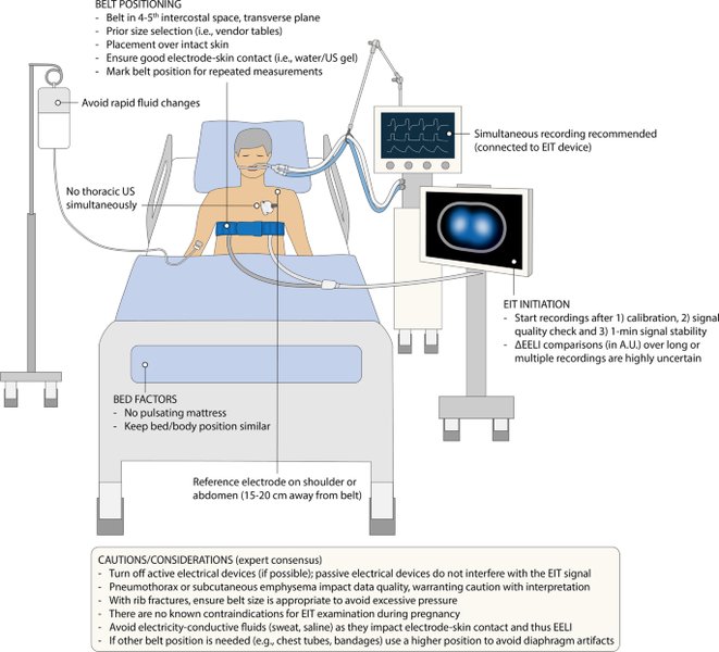
Illustration of EIT electrode belt placement on a patient's chest for lung monitoring.
Beyond Pulmonary Applications
While lung monitoring is a dominant area, EIT's potential extends further:
- Pediatric ECMO Monitoring: EIT is being explored for monitoring pediatric patients on Extracorporeal Membrane Oxygenation (ECMO), where precise and continuous assessment of lung status is vital.
- Brain Activity Imaging: Research is ongoing into using EIT for imaging changes in brain activity, although challenges remain due to the small electrical impedance changes associated with neuronal activity. Functional Electrical Impedance Tomography by Evoke Response (fEITER) is an area of active investigation.
- Tissue Model Imaging: EIT is being used for in-depth imaging of in-vitro tissue models, which is important for research and drug development.
- Industrial Applications: EIT, or more broadly Electrical Tomography, is also finding applications in industrial settings for imaging the distribution of passive electrical properties within objects.
The Future Trajectory of Electrical Impedance Tomography
Innovation on the Horizon
The future of EIT is marked by continuous innovation, particularly in areas like image reconstruction algorithms, hardware development, and expanding clinical applications. The market for Electrical Impedance Tomography systems is projected to grow significantly, from USD 500 million in 2024 to USD 1.2 billion by 2029, indicating strong confidence in its future.
Advancements in Image Reconstruction and AI
One of the most significant areas of development is in overcoming the "ill-posed" nature of the EIT inverse problem. This involves:
- Deep Learning and Neural Networks: Researchers are increasingly applying deep learning techniques and physics-informed neural networks to improve the accuracy and speed of EIT image reconstruction. Optimizing datasets will be key to maximizing the potential of AI in EIT.
- Online Optimization: Methods for online optimization are being developed to allow for more accurate dynamic EIT imaging as data changes in real-time.
- Addressing Inverse Problems: Continued research into advanced mathematical methods, including those used in geophysics for electrical resistance tomography (ERT), will refine EIT's ability to produce clear and reliable images.
Dr. Brian Ring discussing the current research and clinical applications of EIT.
This video provides an excellent overview of how Electrical Impedance Tomography (EIT) is currently being utilized in both research and clinical settings. It delves into the practical applications at the bedside, emphasizing how EIT's non-invasive and real-time capabilities are transforming patient monitoring, particularly in respiratory care. The insights shared by Dr. Ring underscore the technology's immediate impact and its potential to personalize patient care through continuous, dynamic imaging of internal physiological processes.
Hardware Innovation and Wearable Devices
The trend towards miniaturization and portability will continue to make EIT more accessible and versatile:
- Wireless and Wearable Systems: The development of wireless and wearable EIT systems promises to facilitate clinical measurements and open new avenues for home monitoring, enhancing patient comfort and data collection frequency.
- Improved Electrode Technology: Advances in electrode design and placement will enhance signal quality and overall image resolution.
- Multi-frequency EIT: While conventional EIT often uses a single frequency, multi-frequency EIT (MFEIT) is gaining traction. This approach leverages the characteristic that the electrical impedance of human organs and tissues changes at different excitation frequencies, potentially offering richer diagnostic information.
Expanding Clinical Horizons
Beyond current uses, EIT is poised to impact a wider range of medical fields:
- Cardiac Output Monitoring: EIT could play a role in determining cardiac output, offering a less invasive alternative to existing methods.
- Breast Cancer Screening: Research into EIT's potential for screening and monitoring breast cancer is ongoing, leveraging differences in electrical properties between healthy and cancerous tissues.
- Urinary Bladder Monitoring: Wearable devices based on bioimpedance measurements and machine learning are being explored for urinary bladder monitoring.
- Musculoskeletal Applications: Electrical Impedance Myography (EIM), a related technology, is advancing for understanding tissue properties in muscles and related conditions.
Key Conferences and Research Initiatives
Driving EIT Forward
The global EIT community actively collaborates and shares advancements through conferences and research initiatives. These gatherings are crucial for fostering innovation and translating research into clinical practice.
Promotional banner for the ICEBI-EIT-CNIBi 2025 conference.
One of the most anticipated events is the 25th International Conference on Biomedical Applications of Electrical Impedance Tomography (ICEBI-EIT-CNIBi 2025). Scheduled for June 25-27, 2025, in Monterrey, Mexico, this conference will serve as a dynamic forum for researchers, industry professionals, and students. It aims to facilitate the exchange of ideas, knowledge, and research findings in both Bioimpedance and Electrical Impedance Tomography. Key topics for submission include clinical and technological advancements in EIT, as well as new developments in bioimpedance. The deadline for abstract and full paper submissions is February 28, 2025, highlighting the ongoing active research and development in the field.
Beyond this specific conference, various EIT communities and initiatives, such as the European Institute of Innovation & Technology (EIT) and its various KICs (Knowledge and Innovation Communities) like EIT Health, EIT Digital, EIT Manufacturing, and EIT RawMaterials, are actively promoting innovation, entrepreneurship, and skill development across different sectors, some of which indirectly support or utilize technologies related to EIT.
Comparative Overview of EIT and Other Imaging Modalities
A Unique Position in Medical Imaging
To fully appreciate EIT's significance, it's helpful to compare it with more established medical imaging techniques. This table highlights EIT's distinct advantages and current limitations.
| Feature/Modality | Electrical Impedance Tomography (EIT) | X-ray Computed Tomography (CT) | Magnetic Resonance Imaging (MRI) |
|---|---|---|---|
| Imaging Principle | Measures electrical conductivity/resistivity from surface electrodes. | Uses X-rays to create cross-sectional images of internal structures. | Uses strong magnetic fields and radio waves to generate detailed images of organs and soft tissues. |
| Nature of Information | Functional (dynamic physiological changes, e.g., ventilation, perfusion). | Anatomical (structural details, bone, soft tissue density). | Anatomical and some functional (e.g., fMRI for brain activity, perfusion). |
| Radiation Exposure | None (radiation-free). | Significant (ionizing radiation). | None (non-ionizing). |
| Real-Time Monitoring | Yes, continuous bedside monitoring. | No, static images (periodic scans). | No, static images (periodic scans). |
| Portability | High (compact, bedside devices). | Low (large, fixed machines). | Low (very large, fixed machines). |
| Cost | Relatively low. | Moderate to high. | High. |
| Image Resolution | Lower than CT/MRI for anatomical detail, good for dynamic changes. | High. | High. |
| Main Clinical Use | Lung ventilation/perfusion, critical care monitoring. Emerging: brain, hemolysis. | Detailed anatomical imaging, diagnosis of fractures, tumors, internal bleeding. | Soft tissue detail, brain, spinal cord, joints, cancer staging. |
As the table illustrates, EIT carves out a unique niche by offering real-time, non-invasive, and radiation-free functional imaging, making it complementary to, rather than a direct replacement for, established anatomical imaging techniques like CT and MRI.
Challenges and Opportunities
Navigating the Path to Widespread Adoption
Despite its promise, EIT faces certain challenges. The image reconstruction problem, often referred to as the "inverse problem," remains mathematically complex. While progress has been made with deep learning and other algorithms, achieving high spatial resolution comparable to CT or MRI for static images is still an area of active research. Additionally, standardizing EIT data acquisition, processing, and clinical interpretation across different devices and patient populations is crucial for broader clinical adoption.
However, these challenges also present opportunities. The drive for improved image reconstruction algorithms, coupled with advancements in hardware and the increasing demand for continuous, non-invasive monitoring solutions, positions EIT for significant growth. Its low cost, portability, and safety profile make it particularly attractive for applications in resource-limited settings or for long-term patient monitoring where repeated exposure to radiation is undesirable.
Frequently Asked Questions about Electrical Impedance Tomography
Recommended Further Exploration
- Explore the latest advancements in EIT image reconstruction algorithms.
- Discover how EIT is being applied in critical care for organs other than the lungs.
- Learn about the development of portable and wearable EIT devices for continuous home monitoring.
- Investigate the broader impact of artificial intelligence on the future of medical imaging.
References
Last updated May 21, 2025