
Unveiling the Aquatic Lens: A Deep Dive into the Components of a Fish's Eyeball
Exploring the intricate structures that grant fish their unique underwater vision, from the transparent cornea to the light-sensing retina.
The eye of a fish is a marvel of biological engineering, perfectly adapted for vision in an aquatic environment where light behaves very differently than in air. While sharing fundamental structures with terrestrial vertebrates, fish eyes possess unique features enabling them to navigate, hunt, and evade predators in diverse underwater conditions, from sunlit shallows to the darkest depths. Understanding these components reveals how fish perceive their world.
Key Insights into Fish Eye Anatomy
- Underwater Adaptation: Fish eyes are primarily structured for vision in water, meaning the cornea contributes minimally to light refraction, unlike in terrestrial animals. The powerful, spherical lens handles most of the focusing.
- Specialized Lens: The fish lens is typically a rigid, spherical structure that focuses light by moving closer to or further from the retina, rather than changing shape like a human lens. It's composed of dense layers of proteins called crystallins.
- Varied Light Sensitivity: Many fish possess adaptations for low-light conditions, such as a tapetum lucidum, while others can perceive a broad spectrum of colors, sometimes including ultraviolet light.
Core Components of the Fish Eyeball
The fish eyeball, though varying across species, generally comprises several key structures, each playing a vital role in capturing and processing light.
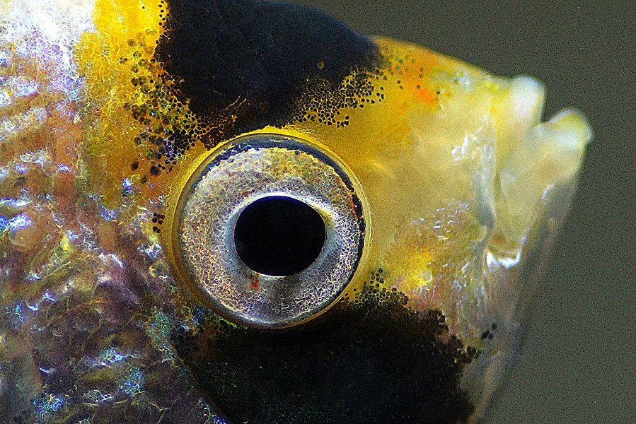
A close-up view of a fish eye, highlighting its prominent, spherical lens adapted for underwater vision.
1. The Cornea: The Outermost Window
The cornea is the transparent, outermost layer of the fish eye. It serves as a protective covering and is the first structure light encounters. Unlike in terrestrial animals where the cornea significantly refracts light due to the air-cornea interface, in fish, the refractive index of water is very similar to that of the cornea. Consequently, the cornea contributes minimally to focusing light. Its typically spherical or dome-shaped structure allows fish to receive light from a very wide field of view, often approaching a 360-degree hemisphere around the eye, crucial for detecting predators and prey from multiple directions.
2. Iris and Pupil: Regulating Light Entry
The iris is the colored part of the eye that surrounds the pupil, an opening that allows light to enter the inner eye. The iris controls the size of the pupil, thereby regulating the amount of light reaching the retina. In many teleost (bony fish) species, the iris is largely immovable, or adjusts very slowly to changes in light intensity, sometimes taking 15 to 20 minutes to adapt. This means sudden bright light can be temporarily blinding. However, elasmobranchs (sharks and rays) possess a more muscular iris, allowing for more effective pupil diameter adjustment. Pupil shapes can vary among species, from circular to slit-like forms.
3. The Lens: The Primary Focusing Element
The lens in a fish eye is a remarkable structure. It is typically very dense, perfectly spherical, and hard. This rigidity is due to its composition of tightly packed protein fibers called crystallins, arranged in concentric, onion-like layers. Because the cornea does little light bending underwater, the spherical lens is responsible for the majority of light refraction needed to focus an image onto the retina.
Focusing Mechanism
Instead of changing shape like the lens in human eyes (accommodation), fish focus by physically moving the entire lens closer to or further from the retina. This movement is controlled by a specialized muscle called the retractor lentis muscle. The lens continues to grow throughout the fish's life, with new layers being added peripherally, similar to the rings of a tree.
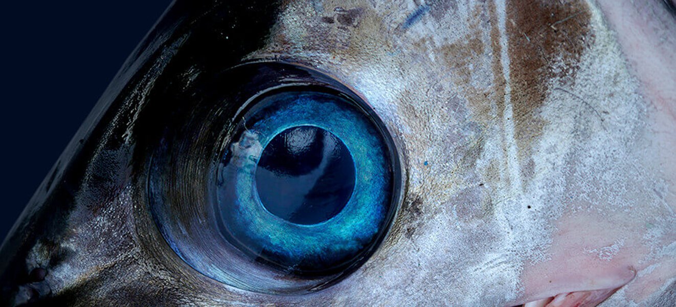
Fish eyes are often positioned laterally, providing a wide field of monocular vision ideal for spotting threats and opportunities in their aquatic habitat.
4. The Retina: Capturing the Image
The retina is the light-sensitive layer lining the back of the eye. It contains photoreceptor cells – rods and cones – that convert light energy into electrical signals. Rods are highly sensitive to low light levels and are primarily responsible for vision in dim conditions, while cones function in brighter light and are responsible for color vision and visual acuity. Many fish species possess excellent color vision, and some can even detect ultraviolet light, which aids in foraging, communication, and mate selection. The light must pass through several neural layers of the retina before reaching the photoreceptors. The retina itself often has a parabolic shape to receive the focused image from the spherical lens.
5. The Optic Nerve: Transmitting Visual Information
The optic nerve is a bundle of nerve fibers that transmits the electrical signals generated by the retina to the brain. The brain then interprets these signals, creating the perception of an image. This connection is vital for processing visual information and enabling appropriate behavioral responses.
6. Internal Fluids and Protective Layers
Aqueous and Vitreous Humor
The fish eye contains two types of fluid. The aqueous humor is a watery fluid found in the anterior chamber, between the cornea and the lens. It helps maintain intraocular pressure and provides nutrients to the cornea and lens. The vitreous humor is a clear, gel-like substance that fills the larger posterior chamber, between the lens and the retina. It helps maintain the eye's shape and optical clarity.
Sclera and Choroid
The sclera is the tough, white, outer protective coat of the eyeball, providing structural support. Internal to the sclera is the choroid, a vascular layer rich in blood vessels that nourishes the retina.
Specialized Adaptations in Fish Eyes
Beyond the fundamental components, many fish species have evolved specialized ocular features to thrive in their specific ecological niches.
Tapetum Lucidum: Enhancing Night Vision
Many nocturnal or deep-sea fish, as well as some elasmobranchs like sharks, possess a tapetum lucidum. This is a reflective layer located behind or within the retina that reflects light that has already passed through the photoreceptor layer back through it a second time. This effectively amplifies the available light, significantly improving vision in low-light or turbid water conditions. The tapetum lucidum is responsible for the "eyeshine" observed in these animals when light is shone into their eyes.
Choroid Rete Mirabile: Oxygen Supply
Some fish, particularly active predators or those living in oxygen-variable environments, have a specialized network of capillaries called the choroid rete mirabile. This structure can secrete oxygen, thereby increasing the oxygen concentration available to the highly metabolic retina. This ensures optimal retinal function even under demanding conditions.
Unique Structures and Behaviors
Nictitating Membrane
While most fish lack eyelids, some sharks possess a nictitating membrane, a translucent third eyelid that can be drawn across the eye for protection, especially during feeding or encounters with other animals.
Four-Eyed Fish (Anableps)
The "four-eyed fish" (genus Anableps) has a remarkable adaptation. Each eye is divided horizontally into two halves, each with its own pupil. This allows the fish to simultaneously see clearly both above and below the water surface as it swims at the waterline, scanning for aerial predators and aquatic prey.
Aphakic Aperture
In some fish species where the pupil size is fixed, an aphakic aperture or space may exist around the lens. This can allow more light to enter the eye, particularly from certain angles, potentially enhancing peripheral vision or sensitivity in low light.
Comparative Visual Capabilities
The visual adaptations of fish vary greatly depending on their habitat and lifestyle. The radar chart below offers a conceptual comparison of key visual parameters between an average shallow-water fish, a deep-sea fish, and a human, highlighting these differences. The scores are relative and illustrative.
This chart illustrates how deep-sea fish excel in low-light sensitivity, while shallow-water fish often have good color and UV perception. Humans, in contrast, have a narrower field of view but relatively good color perception and focus adaptability in air.
Visualizing Fish Eye Anatomy: A Mindmap
The following mindmap provides a hierarchical overview of the primary components of a fish's eyeball and their main roles in the process of vision.
This mindmap illustrates the interconnectedness of the various parts of the fish eye, from the external cornea to the internal retina and optic nerve, highlighting how each contributes to the fish's ability to see underwater.
Summary of Fish Eye Components
The following table provides a concise summary of the main components of a fish's eyeball and their primary functions, emphasizing their adaptations for aquatic life.
| Component | Description & Function |
|---|---|
| Cornea | Transparent outer layer; provides protection; minimal light refraction underwater; contributes to wide field of vision. |
| Iris | Colored muscular diaphragm; controls the size of the pupil to regulate light entry; often slow to adjust in teleosts. |
| Pupil | Opening in the center of the iris; allows light to pass into the lens and onto the retina. |
| Lens | Spherical, rigid, and dense structure; primary focusing element; focuses light by moving closer to or further from the retina; composed of crystallin proteins. |
| Retina | Light-sensitive tissue at the back of the eye; contains photoreceptor cells (rods and cones) that convert light into neural signals. |
| Optic Nerve | Transmits neural signals from the retina to the brain for visual processing. |
| Aqueous Humor | Watery fluid in the anterior chamber (between cornea and lens); maintains intraocular pressure and nourishes avascular tissues. |
| Vitreous Humor | Gel-like substance in the posterior chamber (behind the lens); maintains the shape of the eye and helps transmit light. |
| Sclera | Tough, opaque outer layer; protects the internal structures of the eye. |
| Choroid | Vascular layer between the sclera and retina; provides nutrients and oxygen to the retina. |
| Tapetum Lucidum | (In some species) Reflective layer behind or in the retina; enhances vision in low-light conditions by reflecting light back through photoreceptors. |
| Choroid Rete Mirabile | (In some species) A network of blood vessels; enhances oxygen supply to the retina. |
How Fish Perceive Their World: A Visual Explanation
Understanding the anatomy of a fish's eye is key to appreciating how these creatures perceive their underwater surroundings. The following video provides further insights into fish vision, discussing their unique anatomical features and how they interpret visual information.
This video delves into topics such as color vision in fish, their ability to see in low light, and how the structure of their eyes is perfectly suited for aquatic life. It complements the anatomical details discussed by providing a broader perspective on the functional aspects of fish vision.
Frequently Asked Questions (FAQ)
How do fish focus their eyes?
Unlike humans who change the shape of their lens, fish focus by moving their rigid, spherical lens closer to or further from the retina. This movement is controlled by a special muscle called the retractor lentis.
Can fish see in color?
Yes, many fish species can see in color. Their retinas contain cone cells, which are responsible for color vision. Some fish can even see ultraviolet (UV) light, which is invisible to humans. The extent of color vision varies among different fish species, depending on their habitat and lifestyle.
Why are fish eyes often on the sides of their heads?
The lateral placement of eyes in many fish species provides them with a wide field of monocular vision (each eye sees a different image). This allows them to see almost 360 degrees around their body, which is advantageous for detecting predators and prey. However, this often comes at the cost of a smaller area of binocular vision (where both eyes see the same image), which is important for depth perception.
Do fish have eyelids?
Most fish species do not have eyelids. Since they live in water, their eyes are constantly bathed, which keeps them moist and clean, negating the need for eyelids to perform these functions. Some sharks, however, have a nictitating membrane, a transparent or translucent third eyelid that can cover the eye for protection.
What is the hard sphere found inside a fish eye?
The hard sphere inside a fish's eye is its lens. Unlike the softer, more flexible lens of mammals, a fish's lens is typically very rigid and perfectly spherical. It is made of densely packed proteins called crystallins. Its hardness and shape are crucial for refracting light effectively in an aquatic environment.
Recommended Further Exploration
- Explore the unique visual adaptations of deep-sea fish.
- How does water clarity affect fish vision and behavior?
- Compare the structure and function of fish eyes versus human eyes in detail.
- What role does ultraviolet vision play in fish communication and foraging strategies?
References
Last updated May 14, 2025