
Innovative Diagnostic Techniques in Breast Imaging
Advancements revolutionizing breast cancer detection and diagnosis
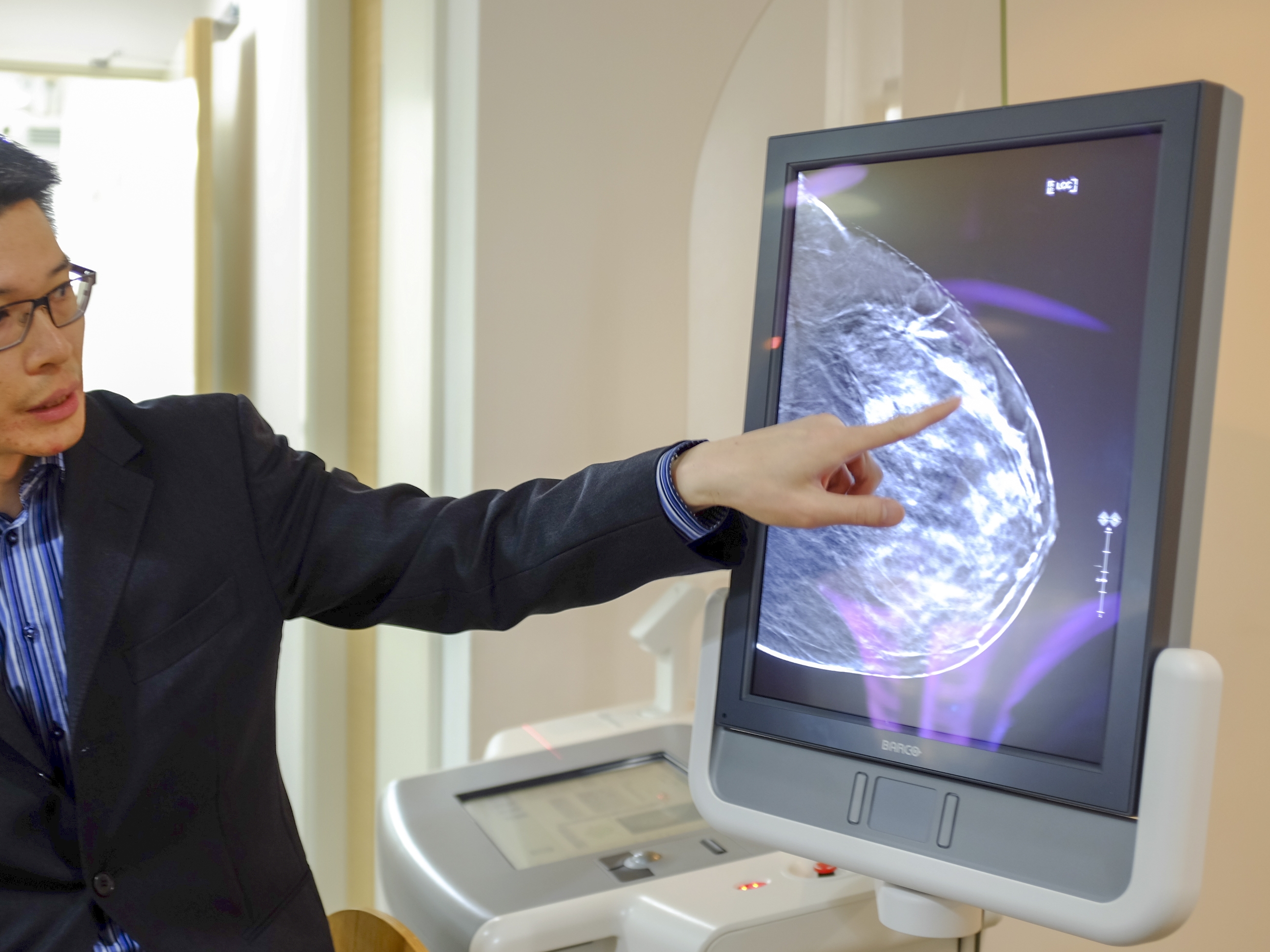
Key Highlights
- Integration of AI and Advanced Imaging: Leveraging artificial intelligence combined with modalities such as digital breast tomosynthesis and contrast-enhanced techniques.
- Enhanced 3D and Molecular Imaging: Utilization of 3D mammography, low-dose positron emission methods, and molecular imaging to improve accuracy and reduce false positives.
- Multimodal and Accessible Systems: Combining different imaging modalities into single platforms that streamline diagnosis and extend services to underserved areas.
Overview of Diagnostic Innovations
The field of breast imaging has witnessed remarkable progress in recent years, propelled by the integration of state-of-the-art technologies and diagnostic methods. These innovative strategies are designed to improve precision in detection, enable earlier cancer diagnosis, and ensure that patient comfort and accessibility are optimally addressed. With developments aimed at enhancing both the sensitivity and specificity of breast imaging techniques, radiologists and healthcare professionals are empowered with tools that significantly improve patient outcomes. The advancements in breast imaging not only translate into improved diagnostic accuracy but also address the needs of high-risk communities and patients with dense breast tissue.
Evolution of Traditional Techniques to Advanced Modalities
Traditional mammography, while crucial in early breast cancer detection, has inherent limitations especially when dealing with dense breast tissue. This limitation has accelerated the development of advanced diagnostic techniques that combine improved imaging modalities with cutting-edge computational analysis. The data-driven revolution, spurred by artificial intelligence, has led to the emergence of more reliable imaging techniques that reduce the incidence of false positives and negatives. With ongoing innovation, healthcare providers are increasingly able to offer more detailed, three-dimensional views of breast tissues, thus enabling a better understanding of abnormalities and potential malignancies.
Artificial Intelligence Integration
One of the most revolutionary additions to breast imaging is the integration of artificial intelligence (AI) algorithms. These systems are designed to analyze mammographic data at a scale and efficiency unattainable by human interpretation alone. AI offers several marked benefits:
- Enhanced accuracy in identifying and characterizing breast lesions.
- Reduction in diagnostic errors by highlighting early signs of malignancy based on pattern recognition.
- Streamlining workflow in radiology departments by pre-screening images and flagging areas of concern.
AI systems utilize vast amounts of imaging data to learn and adapt, thereby improving over time. They have demonstrated particular promise in settings with high patient throughput and in rural or underserved regions where specialist expertise might be less accessible. Furthermore, the growing body of evidence indicates that a combination of AI-augmented analysis with human oversight yields the best results, providing a robust double-check mechanism that increases diagnostic sensitivity and specificity.
Three-Dimensional Mammography (Digital Breast Tomosynthesis)
Digital Breast Tomosynthesis (DBT), commonly referred to as 3D mammography, represents a major evolution in the imaging landscape. By capturing sequential layers or slices of the breast, DBT constructs a comprehensive three-dimensional image. This methodology provides significant advantages over traditional two-dimensional mammograms:
- Improved visualization of overlapping tissues, especially in women with dense breasts.
- Enhanced detection rates of small tumors that may be hidden in standard imaging.
- Reduction in recall rates and false positives, leading to less patient anxiety and unnecessary follow-ups.
The precision of DBT improves radiologist confidence in diagnosing potential malignancies, while also aiding in tailoring personalized treatment and follow-up protocols. The enhanced image clarity helps to determine the extent of cancer spread with greater accuracy, thus supporting more informed clinical decisions.
Contrast-Enhanced Mammography and MRI
Contrast-Enhanced Mammography (CEM) and advanced Magnetic Resonance Imaging (MRI) techniques are pushing the boundaries of diagnostic imaging even further. CEM involves the administration of contrast material, allowing for improved visualization of vascular patterns and tissue perfusion that typically signal malignancy. The advantages of CEM include:
- Comparable sensitivity to MRI for detecting malignancies.
- Shorter imaging times and increased patient accessibility.
- A practical diagnostic alternative in settings where MRI may not be readily available or cost-effective.
Similarly, advanced breast MRI utilizes tailored protocols with lower energy levels and high-definition imaging capabilities. By combining contrast agents with high-resolution imaging, MRI not only supports the detection of early-stage cancers but also provides detailed assessments for high-risk patients. These techniques have found utility particularly in cases where conventional imaging fails to provide a clear diagnosis.
Molecular Imaging Techniques
Molecular imaging techniques are another pillar of the modern diagnostic repertoire. Two techniques stand out:
- Molecular Breast Imaging (MBI): MBI leverages a gamma camera in conjunction with a radioactive tracer to highlight cancerous tissue. Its ability to detect functional changes in tissue, rather than just structural anomalies, makes it particularly effective for patients with dense breasts, where standard mammography may be less sensitive.
- Positron Emission Mammography (PEM): Utilizing a low-dose radioactive tracer, PEM is gaining traction as it correlates closely with the detailed imaging provided by MRI but with a lower risk of false positives. PEM’s high sensitivity in detecting metabolic activity typical of malignant tissues contributes to a prompt and accurate diagnosis.
These molecular imaging techniques provide critical insights into the biochemical behavior of breast tissues, allowing clinicians to detect and monitor cancer progression with unprecedented detail. Their integration into routine screening can enhance early detection, ultimately reducing mortality rates by enabling swift interventions.
Automated and Wearable Imaging Technologies
In addition to the sophisticated imaging modalities already discussed, innovations in automated whole-breast ultrasound (AWBUS) and wearable imaging technologies are making significant strides. AWBUS provides a comprehensive 3D ultrasound imaging of the entire breast, offering an excellent adjunct to mammography for the detection of cancers in dense tissues without the risk associated with ionizing radiation. Meanwhile, research into wearable imaging devices that incorporate ultrasound and infrared imaging is paving the way for more patient-friendly diagnostic tools. These devices are designed to be less invasive and more comfortable, potentially allowing continuous monitoring in high-risk individuals.
These technologies not only broaden the diagnostic armamentarium but also pave the way for more accessible screening programs, particularly in regions where traditional imaging infrastructure may be lacking. The portability and automation inherent in these devices could lead to revolutionary changes in community-level preventive healthcare, ensuring that early detection becomes a staple in remote and rural healthcare environments.
Comprehensive Overview Table
| Technique | Description | Key Benefits |
|---|---|---|
| AI-Powered Imaging | Utilizes artificial intelligence to analyze and interpret imaging data. | Improved accuracy, reduced workload, enhanced pattern recognition. |
| 3D Mammography (DBT) | Creates a three-dimensional image through multiple angled X-rays. | Better visualization of dense tissue, fewer false positives, improved tumor detection. |
| Contrast-Enhanced Mammography | Administers contrast agents to highlight vascular and tissue variations. | High sensitivity similar to MRI, reduced imaging time, broader accessibility. |
| Breast MRI with Contrast | High-definition imaging using contrast for detailed tissue analysis. | Excellent for high-risk groups, detailed evaluation of anomalies, non-invasive. |
| Molecular Breast Imaging (MBI) | Employs gamma cameras with radioactive tracers to detect functional abnormalities. | Effective in dense breasts, functional imaging, complements structural imaging. |
| Low-Dose Positron Emission Mammography (PEM) | Uses low-dose radioactive tracers to detect metabolic activity. | High sensitivity, lower radiation exposure, reduced false positives. |
| Automated Whole-Breast Ultrasound (AWBUS) | Provides a full 3D ultrasound scan of the breast. | Radiation-free, comprehensive coverage, particularly useful in dense tissue. |
| Wearable Imaging Devices | Incorporates ultrasound and infrared technologies for continuous monitoring. | Enhanced patient comfort, portability, potential for remote screening. |
Accessibility and Broader Implications
A major focus of current innovations in breast imaging is ensuring that high-quality diagnostic tools are accessible to all patients, including those in remote and underserved communities. With the advent of AI-powered image analysis and portable imaging solutions, the gap between urban healthcare centers and rural areas is rapidly narrowing. This shift is critically important, as early diagnosis is directly linked with improved treatment outcomes and reduced mortality.
The multipronged approach—integrating AI, advanced imaging modalities, and automated systems—addresses several key challenges:
- Enhancing Diagnostic Accuracy: The synergy between AI and 3D/molecular imaging methods significantly improves the precision of cancer detection, reducing both false positives and negatives.
- Reducing Radiologist Workload: Automated and AI-enhanced systems assist radiologists by pre-processing data and highlighting areas of concern, thereby allowing more efficient and focused evaluations.
- Expanding Access to High-Quality Screening: Portable and wearable imaging devices ensure that even those in geographically challenging areas receive timely and effective diagnostic evaluations.
Moreover, these innovations provide an inclusive diagnostic framework that encompasses a wide demographic, including patients with dense breast tissue or other special needs. This holistic approach is transforming the standard of care by transitioning from a “one-size-fits-all” model to a personalized diagnosis strategy that considers the unique characteristics of every patient.
Technological Synergies and Future Directions
Synergistic Approaches
The integration of various imaging modalities into coherent diagnostic platforms is central to the latest advances in breast imaging. These multimodal systems merge the strengths of conventional mammography, ultrasound, MRI, and molecular imaging into a comprehensive diagnostic tool. Benefits of such integration include:
- Unified Diagnostic Platforms: These platforms allow radiologists to view and analyze data from multiple sources in one interface, enhancing the decision-making process.
- Synchronized Imaging and Analysis: Seamless data sharing between modalities, combined with AI-driven analytics, yields a more complete picture of tissue characteristics.
- Personalization of Care: Integrated systems can adapt imaging strategies based on patient-specific risk profiles and tissue density, ensuring that each woman receives a tailored evaluation.
Additionally, ongoing research continues to refine these methods, aiming to further lower radiation dosages without sacrificing image quality, an important consideration to reduce potential long-term risks. The ongoing evolution of these technologies hints at even more accessible and precise diagnostics in the near future.
Cost Effectiveness and Practical Deployment
Alongside clinical advancements, practical considerations such as cost-effectiveness and ease of deployment play a pivotal role in driving these innovations from research environments into widespread clinical practice. New imaging modalities must not only perform well under strict testing conditions but also integrate seamlessly into everyday medical workflows. To this end, several strategies have been developed:
- Simplified Workflow Integration: New imaging systems are designed to fit into existing clinical infrastructures with minimal disruption, often incorporating automated features that streamline image capture and interpretation.
- Modular System Design: Facilitating upgrades as newer technologies emerge, such modular designs ensure that healthcare institutions can adapt quickly to advancements without overhauling entire systems.
- Remote Diagnostics: Innovations that allow telemedicine integration offer a promising path forward for regions lacking local specialist services, thus democratizing access to high-standard diagnostic care.
By balancing advanced imaging with cost and operational efficiency, clinicians are better equipped to adopt these technologies on a larger scale, ultimately leading to improved patient outcomes and more efficient healthcare delivery.
References
- Latest Innovations in Mammogram Technology for 2025 - InView Imaging
- The Top Trends in Diagnostic Imaging for 2025 - USC Ultrasound
- Breast Imaging Market Size Forecast - PharmiWeb
- U.S. Breast Cancer Screening Diagnostics Forecast - GlobeNewswire
-
Current State of Breast Cancer Diagnosis and Treatment - PubMed Central
Recommended Related Queries
Last updated March 13, 2025