
Unlocking Brain Secrets: The Forefront of Isolating Neuron-Specific Messengers from Blood
Discover cutting-edge techniques to precisely capture neuron-derived extracellular vesicles from plasma for groundbreaking neurological insights.
The isolation of neuron-specific extracellular vesicles (NDEVs) from plasma represents a significant leap forward in neuroscience and diagnostics. These tiny messengers, shed by neurons, travel through the bloodstream carrying a cargo of proteins, lipids, and nucleic acids that mirror the health and pathological state of their parent cells. Accessing this information non-invasively through a blood draw holds immense promise for understanding, diagnosing, and potentially treating a range of neurological and psychiatric disorders. As of May 12, 2025, researchers have developed sophisticated methods to enhance the specificity, purity, and yield of NDEVs from the complex milieu of plasma.
Key Breakthroughs in NDEV Isolation
- Targeted Capture is Paramount: Immunoaffinity methods, employing antibodies against specific neuronal surface proteins such as L1CAM, Neurexin-3 (NRXN3), and ATP1A3, offer the highest precision for selectively isolating neuron-derived EVs from a mixed population.
- Strength in Synergy: Hybrid approaches that combine initial enrichment techniques like Size Exclusion Chromatography (SEC) or refined Ultracentrifugation (UC) with subsequent immuno-isolation steps are proving highly effective in maximizing the purity and yield of NDEVs.
- The Future is Automated and Efficient: Emerging technologies, particularly microfluidics, are paving the way for automated, high-throughput, and gentle NDEV isolation, requiring minimal plasma volumes and reducing processing times.
The Critical Role of Isolating Neuron-Specific EVs
Neuron-derived extracellular vesicles are invaluable because they can cross the blood-brain barrier, offering a real-time molecular snapshot of the central nervous system (CNS) through a peripheral blood sample. Isolating these specific vesicles from the vastly more abundant EVs released by other cell types (e.g., platelets, endothelial cells, immune cells) is crucial. This specificity allows for the identification of biomarkers directly related to neuronal function or dysfunction, which is vital for advancing research into conditions like Alzheimer's disease, Parkinson's disease, multiple sclerosis, and traumatic brain injury.
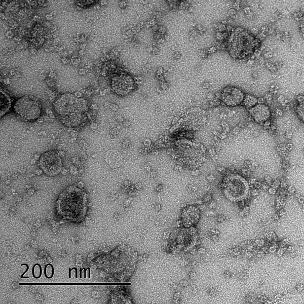
Extracellular vesicles, including those derived from neurons, play a critical role in intercellular communication and are implicated in the pathology of neurodegenerative diseases.
Precision Targeting: Immunoaffinity Isolation Methods
Immunoaffinity-based isolation stands as the most specific approach for capturing NDEVs. This technique relies on the highly selective interaction between antibodies and unique or highly enriched protein markers present on the surface of EVs originating from neurons.
The Principle: Antibodies as Molecular Hooks
This method typically involves incubating plasma, or a pre-enriched EV fraction from plasma, with antibodies that are specific to neuronal surface antigens. These antibodies are often conjugated to a solid support, such as magnetic beads, sepharose resins, or microplate surfaces. When NDEVs encounter their corresponding antibodies, they bind, allowing them to be separated from non-neuronal EVs and other plasma components.
Key Neuronal Surface Markers Driving Specificity
The success of immunoaffinity isolation hinges on the choice of target markers. Several proteins have been identified and validated for their utility in NDEV capture:
- L1 Cell Adhesion Molecule (L1CAM): A well-established marker, L1CAM is abundantly expressed on neuronal EVs and has been widely used for their immunoprecipitation from plasma.
- Neurexin-3 (NRXN3): Recent studies have highlighted NRXN3 as a promising and reliable surface marker for achieving efficient immuno-isolation of NDEVs from both plasma and cerebrospinal fluid (CSF) with high purity.
- ATP1A3: This subunit of the Na+/K+-ATPase is predominantly expressed in neurons, and targeting ATP1A3 on EVs has emerged as a novel strategy for specific NDEV isolation, particularly for diagnostic applications.
- Other potential markers continue to be explored to broaden the toolkit for NDEV capture.
Methodologies and Formats
Immuno-capture can be performed using various formats:
Magnetic Beads: Antibodies are conjugated to magnetic beads. After incubation with the sample, a magnet is used to pull down the bead-EV complexes.
Affinity Chromatography: Antibodies are immobilized on a column matrix. The sample is passed through the column, NDEVs bind, and are later eluted.
Immunoplates: Microtiter plates are coated with capture antibodies, allowing for a more high-throughput approach, especially when combined with automated liquid handling.
Commercial kits, such as ExoSORT, are also available, which utilize neuron-specific antibodies often coupled with magnetic beads to streamline the isolation process.
Synergistic Strategies: Hybrid Isolation Methods
Recognizing that no single method is perfect, researchers increasingly employ hybrid or multi-step approaches to enhance the purity and yield of NDEVs. These strategies typically involve an initial bulk EV enrichment step followed by a more specific immuno-capture stage.
Size First, Specificity Second: SEC-Immunoaffinity
Size Exclusion Chromatography (SEC) is a gentle method that separates EVs from smaller soluble proteins and larger particles based on their hydrodynamic volume. Plasma is passed through a column packed with porous beads; larger EVs elute first, while smaller proteins and contaminants are retarded. The EV-enriched fractions obtained from SEC can then be subjected to immunoaffinity isolation using neuron-specific antibodies. This two-step process effectively reduces background contaminants, improving the efficiency and specificity of the subsequent immuno-capture.
Density and Targeting: UC-Immunoaffinity
Differential Ultracentrifugation (UC), particularly when refined with density gradients (e.g., sucrose or iodixanol gradients), can enrich for EVs based on their characteristic density and size. While UC alone may co-isolate non-EV particles or EVs from other sources, the resulting EV-enriched pellet can serve as a cleaner input for immunoaffinity capture. This combination leverages the concentrating power of UC with the specificity of antibodies.
Advantages of Multi-Step Approaches
Combining methods addresses several challenges:
- It reduces the complexity of the starting material, allowing immunoaffinity reagents to work more efficiently.
- It can improve the overall purity by removing a broader range of contaminants (e.g., lipoproteins, abundant plasma proteins) that might interfere with downstream analyses.
- It helps preserve EV integrity, especially when gentle methods like SEC are used for initial enrichment.
Refining the Classics: Advances in Conventional Techniques
While newer methods offer enhanced specificity, traditional techniques like ultracentrifugation and SEC continue to play a role, often as foundational steps or with modifications that improve their performance for NDEV studies.
Enhanced Ultracentrifugation Protocols
Differential ultracentrifugation (dUC) remains a common method for initial EV pelleting. However, standard dUC can suffer from low purity and potential EV damage. Advances include:
- Density Gradient Ultracentrifugation: Layering the sample on a density gradient (e.g., sucrose or iodixanol) allows EVs to migrate to their buoyant density, separating them more effectively from proteins and lipoproteins.
- Cushion Ultracentrifugation: Pelletting EVs onto a dense cushion (e.g., a small volume of sucrose solution) can help reduce contamination from soluble proteins.
Even with these refinements, UC alone typically yields a mixed EV population, necessitating further specific isolation steps for NDEVs.
Optimized Size Exclusion Chromatography
SEC is valued for its ability to preserve EV integrity and separate EVs from soluble plasma proteins efficiently. Modern SEC protocols utilize optimized column chemistries and dimensions (e.g., commercially available qEV columns) for reproducible EV enrichment from plasma. While SEC alone does not provide neuronal specificity, it is an excellent preparatory step before immunoaffinity capture, significantly improving the quality of the isolated NDEVs by removing contaminants that could interfere with antibody binding or downstream assays.
Visualizing NDEV Isolation Strategies
The following mindmap illustrates the key methodologies and considerations involved in isolating neuron-specific extracellular vesicles from plasma. It provides a hierarchical overview of the main approaches, specific techniques, critical markers, and the overall goals and challenges in this rapidly evolving field.
and specific capture"] id2b["UC + Immunoaffinity"] id2b1["Combines density/size separation
and specific capture"] id3["Refined Conventional Methods"] id3a["Advanced Ultracentrifugation"] id3a1["Density Gradients"] id3a2["Cushion UC"] id3b["Optimized SEC"] id3b1["Pre-enrichment of total EVs"] id4["Emerging Technologies"] id4a["Microfluidics"] id4a1["Automation & High Throughput"] id4a2["Low Sample Volume"] id4a3["Integrated immuno-capture"] id4b["Charge-based Separation"] id4b1["Exploits EV surface charge"] id5["Overall Challenges"] id5a["Standardization of protocols"] id5b["Achieving high Purity & Yield"] id5c["Low abundance of NDEVs in plasma"] id5d["Marker specificity and heterogeneity"] id6["Primary Goals"] id6a["Biomarker Discovery for CNS disorders"] id6b["Non-invasive Diagnostics"] id6c["Understanding Neuronal Pathology"] id6d["Therapeutic Development"]
The Next Wave: Emerging Isolation Technologies
Innovation in NDEV isolation continues, with several promising technologies on the horizon aiming to improve efficiency, automation, and specificity.
Microfluidics: Miniaturization for Maximum Impact
Microfluidic devices, or "lab-on-a-chip" systems, offer significant advantages for EV isolation. These platforms can integrate multiple separation principles (e.g., size-based filtration, acoustic separation, immunoaffinity capture) into a single, automated workflow. Benefits include:
- Minimal Sample Volume: Often requiring only microliters of plasma.
- Reduced Processing Time: Isolation can be completed much faster than conventional methods.
- High Throughput Potential: Suitable for processing multiple samples in parallel.
- Gentle Handling: Shear forces can be precisely controlled, minimizing EV damage.
Microfluidic chips functionalized with neuron-specific antibodies are being developed to directly capture NDEVs from raw or minimally processed plasma, streamlining the entire isolation process.
Harnessing Charge: Physicochemical Separation
EVs generally possess a net negative surface charge. Charge-based isolation techniques exploit this property using ion-exchange chromatography or charged bead materials to capture EVs or deplete oppositely charged contaminants. While typically not specific to neuronal origin on its own, this approach can serve as an effective pre-enrichment step, complementing immunoaffinity or other targeted methods by removing bulk proteins and other impurities from plasma.
Comparative Analysis of NDEV Isolation Methods
Choosing the optimal method for isolating neuron-specific EVs depends on various factors, including the specific research question, available resources, required purity, and downstream applications. The radar chart below provides a comparative overview of different isolation strategies based on key performance criteria. The scores are indicative, reflecting general trends discussed in the literature.
This chart highlights that hybrid methods like SEC followed by immunoaffinity often provide a good balance of specificity, purity, and gentleness, while emerging microfluidic techniques promise high throughput and automation. The choice ultimately depends on the specific experimental needs and trade-offs one is willing to make.
Summary Table of Isolation Methods
The following table provides a concise summary of the primary methods used for isolating neuron-specific extracellular vesicles from plasma, outlining their principles, key markers or features, advantages, and common disadvantages.
| Method Category | Principle | Key Neuron-Specific Markers/Features | Advantages | Disadvantages |
|---|---|---|---|---|
| Immunoaffinity (Standalone) | Antibody-antigen binding to neuron-specific surface proteins. | L1CAM, NRXN3, ATP1A3 | Highest specificity; yields relatively pure NDEV population; direct targeting. | Higher cost (antibodies); potential for marker heterogeneity leading to incomplete capture; yield can be variable. |
| SEC + Immunoaffinity | Initial size-based separation (SEC) to enrich EVs and remove soluble proteins, followed by specific antibody capture. | L1CAM, NRXN3, ATP1A3 (post-SEC) | Good purity and EV integrity; reduces contaminants before specific capture; improves immunoaffinity efficiency. | Multi-step process; potential for sample loss at each step; SEC can dilute samples. |
| UC + Immunoaffinity | Initial EV concentration/enrichment by ultracentrifugation, followed by specific antibody capture. | L1CAM, NRXN3, ATP1A3 (post-UC) | Effectively concentrates EVs from larger volumes before specific capture. | Time-consuming; UC can cause EV aggregation or damage; potential co-precipitation of contaminants with EVs. |
| Advanced Ultracentrifugation (e.g., Density Gradient) | Separation based on buoyant density and size through sequential centrifugation or within a density gradient. | General EV enrichment (not neuron-specific alone). | Well-established; scalable; can separate EVs from some lipoproteins and protein aggregates. | Lower specificity for NDEVs; co-isolates EVs from all cellular origins; can be harsh on vesicles. |
| Size Exclusion Chromatography (SEC) | Separation based on hydrodynamic volume (molecular sieving); larger particles (EVs) elute before smaller ones (proteins). | General EV enrichment (not neuron-specific alone). | Gentle, preserves EV integrity and function; effectively removes soluble proteins; relatively fast. | Lower specificity for NDEVs alone; potential for some overlap with similar-sized particles (e.g., large lipoproteins); can dilute sample. |
| Microfluidics | Miniaturized separation using various principles (e.g., size-based, acoustic, immunoaffinity) on a chip. | Can integrate immuno-tags (L1CAM, etc.) for specificity. | Fast processing; automation potential; requires very small sample volumes; high-throughput capability; gentle handling. | Still an emerging field; standardization can be challenging; initial setup costs may be higher; some designs may be prone to clogging. |
| Charge-Based Isolation | Exploits the net negative surface charge of EVs for separation using ion-exchange materials. | General EV enrichment (not neuron-specific alone). | Rapid; can remove certain types of impurities (e.g., some proteins). | Lower specificity for NDEVs; may require further purification for specific applications. |
Insights from the Field: EV Isolation Techniques
Understanding the nuances of extracellular vesicle isolation is crucial for researchers. The video below from Izon Science discusses general considerations and challenges when isolating EVs from plasma. While not exclusively focused on neuron-specific EVs, the principles of handling plasma, removing contaminants, and choosing appropriate general EV isolation methods (like SEC, which is often a precursor to neuron-specific immunoaffinity steps) are highly relevant. These foundational techniques are often adapted and combined with specific targeting strategies to successfully isolate NDEVs.
General strategies for isolating extracellular vesicles from plasma, providing context for more specialized neuron-specific methods.
The isolation of neuron-specific EVs from plasma involves careful pre-analytical plasma processing to remove platelets and cellular debris, often followed by one or more of the general EV isolation techniques discussed, such as SEC or optimized UC, to enrich for total EVs. The final, critical step then involves immunoaffinity capture using antibodies against validated neuronal surface markers to achieve the desired specificity. Each step must be optimized to maximize yield and purity while preserving the integrity of the NDEVs and their valuable molecular cargo for downstream analysis.
Frequently Asked Questions (FAQ)
What are extracellular vesicles (EVs)? +
Extracellular vesicles (EVs) are nano-sized, membrane-bound particles released by virtually all cell types, including neurons. They act as intercellular messengers, transporting a diverse cargo of proteins, lipids, and nucleic acids (like mRNA, miRNA) between cells. Common types of EVs include exosomes, microvesicles, and apoptotic bodies, distinguished by their biogenesis, size, and composition.
Why is it important to isolate neuron-specific EVs from plasma? +
Plasma contains a heterogeneous mixture of EVs from various cell types. Isolating neuron-specific EVs (NDEVs) allows researchers to access molecular information directly from the central nervous system (CNS) through a minimally invasive blood draw. This is crucial for discovering biomarkers related to neuronal health or disease, understanding brain pathology, and developing targeted diagnostics for neurological disorders like Alzheimer's, Parkinson's, or multiple sclerosis, without needing direct access to brain tissue or CSF.
What are the main challenges in isolating neuron-specific EVs? +
Key challenges include:
1. Low Abundance: NDEVs are typically present in much lower concentrations in plasma compared to EVs from other sources like platelets or endothelial cells.
2. Specificity of Markers: Identifying surface markers that are truly exclusive to neuronal EVs or highly enriched on them is ongoing research. Some markers may show expression on other cell types at low levels.
3. Purity: Separating NDEVs from other EVs and contaminants like lipoproteins and abundant plasma proteins requires highly efficient methods.
4. Yield: Achieving sufficient yield for downstream analysis, especially from small plasma volumes, can be difficult.
5. Standardization: Lack of standardized protocols across different labs can make it hard to compare results.
How are isolated neuron-specific EVs characterized? +
Characterization ensures the isolated particles are indeed EVs and are of neuronal origin. Common techniques include:
- Nanoparticle Tracking Analysis (NTA): To determine size distribution and concentration.
- Transmission Electron Microscopy (TEM) or Cryo-EM: To visualize EV morphology (cup-shaped or spherical).
- Western Blotting or Flow Cytometry: To detect general EV markers (e.g., CD9, CD63, CD81, ALIX, TSG101) and specific neuronal markers (e.g., L1CAM, NCAM, NRXN3, ATP1A3, synaptophysin). The absence of non-EV contaminant markers (e.g., apolipoproteins for lipoproteins, albumin for plasma proteins) is also important.
- Proteomic, Transcriptomic, or Lipidomic Analysis: To profile the molecular cargo of the NDEVs.
What are the downstream applications of isolated neuron-specific EVs? +
Isolated NDEVs have numerous applications:
1. Biomarker Discovery: Identifying proteins, RNA, or lipids within NDEVs that can serve as diagnostic, prognostic, or predictive biomarkers for neurological diseases.
2. Disease Monitoring: Tracking changes in NDEV cargo over time to monitor disease progression or response to therapy.
3. Understanding Pathogenesis: Studying the molecular contents of NDEVs can provide insights into the mechanisms underlying brain disorders.
4. Drug Delivery: Engineered EVs are being explored as potential vehicles for delivering therapeutic agents across the blood-brain barrier to target neurons.
5. Personalized Medicine: NDEV profiles could help tailor treatments for individuals with neurological conditions.
Recommended Further Exploration
- What are the specific advantages of using NRXN3 as a marker for neuron-derived EV isolation compared to L1CAM?
- How do microfluidic devices integrate immunoaffinity capture for isolating neuron-specific EVs?
- What are the challenges in standardizing protocols for neuron-specific EV isolation for clinical applications?
- How can the molecular cargo of neuron-specific EVs be analyzed for biomarker discovery in Alzheimer's disease?
References
Last updated May 12, 2025