
Unlocking Neurological Insights: Cutting-Edge Methods for Isolating Neuron-Specific Extracellular Vesicles from Plasma
Explore the advanced techniques revolutionizing biomarker discovery and disease monitoring through targeted EV isolation.
Key Advances in Neuron-Specific EV Isolation
- Immunoaffinity Capture: Leveraging highly specific antibodies to target neuronal surface markers like L1CAM, NRXN3, and ATP1A3, enabling precise isolation of neuron-derived EVs.
- Hybrid Approaches for Enhanced Purity: Combining initial bulk EV isolation methods such as Size Exclusion Chromatography (SEC) or ultracentrifugation with subsequent immunoaffinity capture to achieve superior purity and specificity.
- Emerging Microfluidics and Rapid Filtration: Pioneering technologies like microfluidic devices, charge-based electrokinetic mesh filtration (ExoFilter), and osmosis-driven filtration (EV-Osmoprocessor) are paving the way for faster, more automated, and scalable isolation.
The isolation of neuron-specific extracellular vesicles (nEVs) from plasma represents a significant frontier in neuroscience, offering a non-invasive window into the brain's health and disease states. As of June 2025, the field has seen remarkable advancements, moving towards more specific, pure, and efficient isolation techniques. These innovations are critical for unlocking the full potential of nEVs as biomarkers for neurodegenerative disorders, brain injury, and other neurological conditions. The primary challenge lies in separating these minute vesicles, which carry neuronal cargo (proteins, lipids, nucleic acids), from the vast and heterogeneous population of EVs released by other cell types in plasma, as well as from abundant plasma proteins and lipoproteins. Current state-of-the-art methods often combine foundational EV isolation principles with highly targeted strategies, enhancing both yield and specificity.
Targeted Isolation through Immunoaffinity Capture
Immunoaffinity capture is a cornerstone of neuron-specific EV isolation, relying on the highly selective binding of antibodies to unique surface markers present on neuronal EVs. This method allows researchers to 'pull out' nEVs from the complex plasma milieu, even when they are present in very small quantities. The specificity of this approach is paramount for ensuring that the isolated vesicles truly reflect neuronal pathology rather than systemic inflammation or other confounding factors.
Key Neuronal Surface Markers
The success of immunoaffinity capture hinges on the identification and validation of robust neuron-specific surface markers. Several key proteins have emerged as leading candidates for this purpose:
- L1CAM (L1 Cell Adhesion Molecule): L1CAM has been extensively utilized as a neuronal marker due to its enrichment on EV surfaces of neuronal origin. It facilitates cell adhesion and signaling crucial for neural development and function. Protocols often employ L1CAM antibodies to immunoprecipitate neuron-derived EVs (e.g., L1CAM+ EVs) after an initial broader EV isolation step.
- NRXN3 (Neurexin 3): More recently, Neurexin 3 has been identified and validated as a neuron-specific transmembrane protein present on EVs. Studies have shown its utility in immuno-isolation from both plasma and cerebrospinal fluid (CSF), offering an alternative or complementary target to L1CAM.
- ATP1A3 (Neuronal Sodium/Potassium ATPase α3 subunit): This protein, abundantly expressed in EVs from induced human neurons, brain, CSF, and plasma, is another promising target for neuron-specific EV isolation. ATP1A3 plays a vital role in maintaining neuronal membrane potential and is a crucial marker for capturing distinct neuronal EV populations.
- NCAM (Neural Cell Adhesion Molecule): While less commonly used than L1CAM, NCAM also serves as an alternative neuronal marker, contributing to neuronal plasticity and connectivity.
These markers enable highly specific isolation, allowing for subsequent downstream analyses such as proteomic profiling and RNA sequencing, which can reveal crucial insights into neurodegenerative diseases like Alzheimer's and Parkinson's.
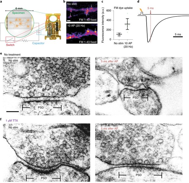
An illustration depicting synaptic vesicles, which are a type of extracellular vesicle, involved in neuronal communication.
Hybrid Approaches for Enhanced Purity and Yield
To overcome the limitations of single-method isolation (e.g., contamination from co-isolated proteins or low yield), many cutting-edge protocols now advocate for combining multiple techniques. These hybrid approaches leverage the strengths of different methods to achieve superior purity, specificity, and yield of neuron-specific EVs from plasma.
Size Exclusion Chromatography (SEC) Coupled with Immunoprecipitation
SEC is increasingly recognized for its ability to produce highly pure EV samples with minimal protein contamination. When combined with immunoaffinity capture, SEC offers a powerful strategy:
- Principle: Plasma is first fractionated by SEC columns (e.g., SmartSEC system) to separate EVs from smaller soluble proteins and lipoproteins based on size. This preliminary step significantly reduces background contamination.
- Integration: The EV-enriched fractions from SEC are then subjected to immunoaffinity capture using beads conjugated with antibodies against neuron-specific markers like L1CAM or NRXN3. This sequential approach ensures that only the neuronal subset of EVs is isolated from an already cleaner sample.
- Advantages: This combination yields purer EV fractions, making them more suitable for sensitive downstream analyses like proteomics or RNA sequencing. It also maintains EV integrity due to the gentle nature of SEC.
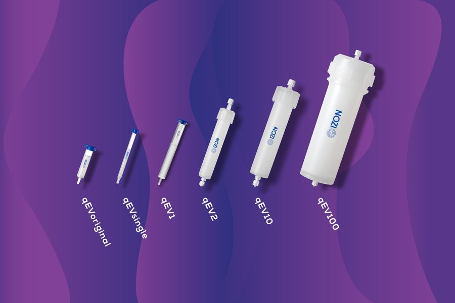
A depiction of a Size Exclusion Chromatography (SEC) column used for extracellular vesicle isolation, highlighting its efficiency in separating EVs by size.
Differential Ultracentrifugation (dUC) with Immuno-Isolation
Despite the emergence of newer techniques, dUC remains a foundational method for EV isolation, often referred to as the "gold standard." Its integration with immuno-isolation offers a robust, albeit time-consuming, approach:
- Principle: Plasma samples undergo a series of centrifugation steps at increasing speeds to sequentially remove cells, cellular debris, and larger vesicles, culminating in ultracentrifugation to pellet EVs.
- Refinement: To achieve neuron specificity, the total EV pellet obtained from dUC is then subjected to immunoaffinity capture using magnetic beads functionalized with neuron-specific antibodies. Density gradient centrifugation can also be incorporated to further refine EV populations based on buoyant density and remove co-isolated proteins.
- Advantages: dUC is scalable and widely available, capable of handling larger sample volumes. When combined with immuno-isolation, it provides high purity for targeted neuronal EVs.
- Limitations: This method is often criticized for being time-intensive, potentially causing EV aggregation, and requiring specialized equipment. However, its continued use, especially as a preliminary step, underscores its effectiveness in yielding high-purity EV samples.
This video provides an insightful lecture on ultracentrifugation for isolating extracellular vesicles, a fundamental method often used as a precursor to more specific neuron-derived EV isolation techniques from plasma. It explains the principles and practical considerations, emphasizing the importance of this method in EV research.
Emerging Technologies and Future Directions
The quest for faster, more precise, and scalable neuron-specific EV isolation methods has led to the development of several innovative technologies. These emerging approaches promise to revolutionize clinical diagnostics and research by enabling rapid and high-throughput analysis of nEVs.
Microfluidics-Based Technologies
Microfluidic devices offer precise control over fluid dynamics and surface chemistry, enabling rapid EV isolation from small sample volumes. Some notable advancements include:
- On-Chip Capture: Researchers are developing microfluidic chips with surface-immobilized neuron-specific antibodies for direct, on-chip capture of neuron-derived EVs from plasma. This streamlines the process, reducing sample handling and processing time.
- Advantages: These systems offer high efficiency, minimal sample and reagent consumption, and the potential for automation, making them ideal for high-throughput clinical applications.
Rapid Filtration Techniques
Novel filtration-based methods are also gaining traction for their speed and scalability:
- Charge-Based Electrokinetic Mesh Filtration (ExoFilter): This ultrafast technique uses electrostatic forces to filter EVs, offering a high-throughput solution without the need for ultracentrifugation. It has shown promise in isolating EVs from various biofluids, including plasma, and can be adapted for neuron-specific capture.
- Osmosis-Driven Filtration (EV-Osmoprocessor): The EV-Osmoprocessor leverages osmosis for rapid and efficient EV enrichment. This novel device focuses on concentrating EVs quickly, significantly reducing processing time and offering potential for integration with immunoaffinity methods for neuron-specific isolation.
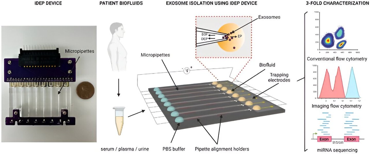
An image illustrating the concept of rapid purification and characterization of circulating small extracellular vesicles, reflecting advancements in filtration technologies.
Comparative Analysis of Isolation Methods
To provide a clearer understanding of the trade-offs involved in selecting an isolation method, the following radar chart illustrates a subjective assessment of various techniques based on key performance indicators for neuron-specific EV isolation. This chart is based on the consensus and prevalent ideas from the provided answers, reflecting general trends and reported advantages/limitations.
This radar chart provides a visual comparison of different isolation methods based on their perceived strengths across various criteria. Purity refers to the absence of contaminating proteins and non-EV particles. Yield indicates the quantity of EVs recovered. Specificity measures the accuracy in isolating only neuron-derived EVs. Speed reflects the processing time. Cost encompasses equipment and reagent expenses. Scalability relates to the potential for high-throughput processing. Hybrid methods like SEC + Immunoaffinity Capture often rank high in both purity and specificity, while emerging technologies excel in speed and scalability, indicating their future potential for clinical translation.
Practical Considerations and Pretreatment
Beyond the primary isolation techniques, several practical considerations are crucial for optimizing the isolation of neuron-specific EVs from plasma. These include sample pretreatment and the selection of appropriate controls.
Sample Pretreatment
The quality of the starting plasma sample significantly impacts the purity and yield of isolated nEVs. Key pretreatment steps include:
- Platelet-Free Plasma: Using platelet-free plasma as the starting material is essential to reduce contamination from platelet-derived EVs, which are highly abundant in blood and can obscure signals from less numerous nEVs.
- Anticoagulants: The choice of anticoagulant (e.g., EDTA, citrate) and processing conditions can influence EV integrity and yield.
- Frozen vs. Fresh Plasma: Studies have shown no significant difference in EV isolation between frozen and fresh plasma samples, which is vital for studies utilizing biobanked samples. However, consistent handling and storage protocols are critical.
- Additives: The evaluation of additives like rabbit thromboplastin (rTP) versus human recombinant thromboplastin (huTP) for plasma pretreatment is an ongoing area of research, with careful consideration of their impact on EV characteristics.
Validation and Controls
Rigorous validation of isolated EVs is paramount to ensure their neuronal origin and suitability for downstream applications:
- Nanoparticle Tracking Analysis (NTA): Used to quantify EV concentration and size distribution.
- Electron Microscopy: Provides visual confirmation of EV morphology and integrity.
- Proteomic Profiling (LC-MS/MS): Confirms the presence of neuron-specific proteins and assesses overall purity.
- Controls: Incorporating controls for non-neuronal EV contamination and non-specific antibody binding is essential to ensure the specificity and reliability of the isolation.
Key Aspects in Neuron-Specific EV Isolation
The following mind map provides a structured overview of the critical components and considerations in isolating neuron-specific extracellular vesicles from plasma, integrating the various methods, markers, and practical aspects discussed.
This mind map visually organizes the complex landscape of neuron-specific EV isolation, illustrating the interconnectedness of various methods, the importance of specific markers, and the overarching goal of utilizing these vesicles for neurological biomarker discovery.
Summary of Key Markers and Their Applications
The choice of neuronal marker is crucial for the success of immunoaffinity-based isolation methods. The table below summarizes the key markers currently in use, their descriptions, and notes on their application and known characteristics.
| Marker | Description | Usage in Isolation | Notes |
|---|---|---|---|
| L1CAM | Neuronal cell adhesion molecule involved in neural development and function. | Commonly used immunocapture target for neuron-derived EVs. | Widely adopted; some debate exists regarding its pan-EV association, but remains a strong neuronal marker. |
| NRXN3 | Neurexin 3, a neuron-specific transmembrane protein involved in synaptic adhesion. | Recently validated for immuno-isolation of neuron-specific EVs from plasma and CSF. | Offers improved specificity; provides a valuable alternative to L1CAM. |
| ATP1A3 | Neuronal sodium/potassium ATPase alpha-3 subunit. | New target for isolating neuron-specific EVs; highly expressed in neuronal EVs. | Promising marker demonstrated in plasma, CSF, and brain tissue. |
| NCAM | Neural cell adhesion molecule involved in cell-cell recognition and adhesion. | Alternative neuronal marker for EV isolation. | Less commonly used than L1CAM, but still a relevant neuronal surface protein. |
This table highlights the diversity of neuronal markers available and their specific roles in refining the isolation of neuron-specific EVs. The ongoing discovery and validation of new markers continue to enhance the precision of these methods.
Frequently Asked Questions (FAQ)
What are neuron-specific extracellular vesicles (nEVs)?
Why is it challenging to isolate neuron-specific EVs from plasma?
What are the main advantages of combining multiple isolation methods?
What are the potential applications of isolated neuron-specific EVs?
Conclusion
The field of neuron-specific extracellular vesicle isolation from plasma is undergoing a rapid evolution, driven by the profound potential of these vesicles as non-invasive biomarkers for neurological conditions. The latest advancements emphasize hybrid approaches that combine initial bulk EV enrichment (e.g., SEC or differential ultracentrifugation) with highly specific immunoaffinity capture using neuronal surface markers such as L1CAM, NRXN3, and ATP1A3. These integrated strategies significantly improve the purity and specificity of isolated nEVs, making them more suitable for detailed molecular analysis. Furthermore, emerging technologies like microfluidics and rapid filtration systems promise to enhance the speed, automation, and scalability of these isolation processes, paving the way for broader clinical applicability. As research continues to refine these methods and validate new neuronal markers, neuron-derived EVs are set to become an indispensable tool in neurological diagnostics, therapeutic monitoring, and our understanding of brain health.
Recommended Further Exploration
- How are extracellular vesicles utilized as biomarkers for neurodegenerative diseases?
- What are the current challenges in exosome isolation from complex biofluids?
- What is the future potential of microfluidic devices in extracellular vesicle diagnostics?
- Why is standardization of extracellular vesicle isolation protocols crucial for clinical translation?