
Understanding the Cervical Cross Section of the Mesial Root of the Lower First Molar
A Comprehensive Analysis of Morphological Characteristics and Clinical Implications
Key Takeaways
- Shape Variability: The cervical cross section of the mesial root exhibits a range of shapes, primarily ovoid, elliptical, or kidney bean-like, influenced by anatomical variations and age-related changes.
- Root Canal Configuration: Typically housing two root canals, the mesial root's morphology facilitates the close relationship or connection between these canals, impacting endodontic treatment approaches.
- Clinical Relevance: Understanding the intricate morphology of the mesial root is crucial for effective dental procedures, including root canal therapy, to ensure comprehensive treatment and prevent procedural complications.
Introduction
The mesial root of the lower first molar is a pivotal structure in dental anatomy, playing a critical role in mastication and overall dental health. Its cervical cross section provides valuable insights into its morphology, influencing various dental procedures, particularly endodontic treatments. This comprehensive analysis delves into the shape, structural variations, and clinical implications of the cervical cross section of the mesial root, synthesizing information from authoritative dental sources.
Morphological Characteristics
General Shape
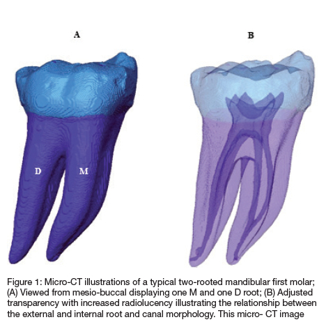
The cervical cross section of the mesial root of the lower first molar typically presents an ovoid or elliptical shape. This elongated oval configuration allows for a broader buccolingual dimension compared to the mesiodistal axis, accommodating the internal root canal system efficiently.
Ovoid and Elliptical Configurations
The predominance of ovoid or elliptical shapes facilitates the accommodation of dual root canals within the mesial root. This shape is not only aesthetically pertinent but also functionally significant, as it influences the access and navigation during endodontic procedures.
Kidney Bean-Like Morphology
Some anatomical studies describe the cervical cross section as resembling a kidney bean. This analogy underscores the presence of a concave aspect on one side, often the distal side, contributing to a non-uniform contour. This concavity may house the isthmus connecting the two canals, reflecting the root's flattened or ribbon-like nature.
Age-Related Shape Modifications
With advancing age, the cervical cross section of the mesial root may exhibit a transition from a more angular or triangular shape to a rounded morphology. This alteration is attributed to the continuous remodeling of the dentin and other structural changes within the root, enhancing the smoothness and reducing the sharpness of the cross-sectional outline.
Internal Structure and Root Canal System
Root Canal Configuration
Typically, the mesial root houses two root canals: one buccal and one lingual. These canals are often in close proximity, sometimes exhibiting an isthmus or an anastomosis, which can bridge the two canals. The intimate relationship necessitates meticulous attention during endodontic procedures to ensure complete debridement and obturation.
Isthmus Presence
The isthmus, a narrow band of dentin, may connect the buccal and lingual canals. Its presence can complicate root canal treatments, as it may harbor remnants of pulp tissue or bacteria, potentially leading to treatment failures if not adequately addressed.
Canal Curvatures and Variations
Variations in canal curvature and morphology are common. Some individuals may present with additional canals or unusual curvatures, emphasizing the need for individualized treatment planning based on thorough radiographic and clinical assessments.
Surface Features and Anatomical Considerations
The mesial root's surface is generally broader buccolingually compared to mesiodistally. Prominent root depressions may be present on both mesial and distal surfaces, contributing to the overall cross-sectional shape. These depressions can influence the tooth's stability and its response to orthodontic forces.
Longitudinal Depressions
Shallow longitudinal depressions on the mesial surface serve as anatomical landmarks, aiding in the identification of root canals and guiding endodontic access and instrumentation.
Broader Buccolingual Dimension
The buccolingual breadth provides ample space for the dental structures and facilitates the accommodation of the root canal system. This dimension is vital for maintaining the tooth's integrity and resistance against lateral forces during mastication.
Clinical Implications
Endodontic Treatment Considerations
The intricate morphology of the mesial root's cervical cross section demands precise endodontic techniques. Understanding the root's shape aids in creating adequate access cavities, locating all canal orifices, and effectively negotiating canal curvatures. Failure to recognize the root's morphology can result in missed canals, ledging, or perforations, compromising the treatment's success.
Access Cavity Design
A well-designed access cavity, considering the root's shape, ensures optimal visibility and accessibility to all canals. The elongated or kidney bean-like shape necessitates strategic angulation and extension of the access to encompass the canal system comprehensively.
Instrumentation and Obturation
Appropriate instrumentation techniques, tailored to the root's curvature and canal configuration, are essential for thorough cleaning and shaping. Similarly, obturation materials and methods must adapt to the canals' shape to achieve a hermetic seal, preventing bacterial ingress and reinfection.
Preventive and Restorative Measures
Knowledge of the mesial root's cross-sectional anatomy informs preventive strategies, such as sealant placement and occlusal adjustments, to mitigate carious progression and structural weaknesses. Restorative procedures, including crown fabrication, benefit from this understanding to ensure proper fit and distribution of occlusal forces.
Caries Prevention
Effective caries prevention mandates a comprehensive approach, considering the root's anatomical features that may predispose it to decay. Sealants and fluoride applications should be strategically placed to fortify vulnerable areas identified through anatomical assessments.
Occlusal Force Distribution
An accurate restoration ensures equitable distribution of occlusal forces, reducing the risk of root fractures or marginal gaps. The broader buccolingual dimension of the mesial root necessitates restorations that accommodate its shape without compromising structural integrity.
Anatomical Variations Among Individuals
Population-Based Morphological Differences
Anatomical variations in the mesial root's cross-sectional shape can occur across different populations. Factors such as genetics, age, and developmental influences contribute to these differences, affecting the prevalence of certain root configurations and root canal systems.
Genetic Influences
Genetic predispositions can result in diverse root morphologies, influencing the shape and number of canals. Understanding these genetic factors assists clinicians in anticipating anatomical complexities during treatment planning.
Age-Related Changes
As individuals age, dental structures undergo remodeling, leading to alterations in root shape and canal morphology. These changes necessitate adaptive treatment approaches to accommodate the evolving anatomy, ensuring effective dental care across the lifespan.
Diagnostic Tools and Techniques
Radiographic Analysis
Advanced radiographic techniques, including periapical and cone-beam computed tomography (CBCT), offer detailed visualization of the mesial root's cross-sectional anatomy. These imaging modalities facilitate accurate diagnosis, treatment planning, and identification of anatomical variations or pathologies.
Cone-Beam Computed Tomography (CBCT)
CBCT provides three-dimensional imaging of dental structures, enabling precise assessment of root morphology, canal pathways, and potential anomalies. Its high-resolution images support meticulous treatment planning and enhance the clinician's ability to address complex anatomical scenarios.
Digital Radiography
Digital radiography offers rapid, high-quality images with the advantage of digital manipulation for enhanced visualization. This technology aids in identifying root shapes, canal configurations, and detecting periapical pathologies with minimal radiation exposure compared to traditional radiography.
Intraoral Imaging Techniques
Intraoral cameras and magnification tools, such as dental operating microscopes, augment the clinician's ability to observe the mesial root's cervical cross section directly. These tools enhance visual acuity, facilitating the detection of subtle anatomical features and improving procedural accuracy.
Dental Operating Microscopes
Microscopes provide magnified, illuminated views of the dental anatomy, enabling precise identification of canal orifices, anatomical variations, and restorative margins. Their integration into clinical practice elevates the standard of care, particularly in intricate procedures like endodontics.
Intraoral Cameras
Intraoral cameras capture real-time images of the dental structures, offering both the clinician and patient a clear view of the treatment area. This transparency facilitates better communication, diagnosis, and procedural execution, especially when navigating complex root anatomies.
Illustrative Examples and Comparative Analysis
Comparative Morphology
Comparing the mesial root's cervical cross section across different molars and populations highlights significant morphological variations. Such comparisons aid in understanding the range of normal anatomical presentations and the implications for dental treatments.
Mandibular vs. Maxillary Molars
While mandibular first molars typically exhibit an ovoid or kidney bean-shaped cervical cross section, maxillary molars may display different configurations based on their distinct functional and anatomical roles. Understanding these differences is crucial for accurate diagnosis and treatment across tooth types.
Population-Specific Variations
Studies indicate that certain populations may exhibit prevalence of particular root shapes or canal configurations, influenced by genetic and environmental factors. Recognizing these trends enhances the clinician's preparedness to encounter and manage diverse anatomical presentations.
Clinical Case Studies
Analyzing clinical cases where the mesial root's cross section deviates from the norm provides practical insights into managing complex anatomical scenarios. These cases underscore the importance of tailored treatment approaches and the utility of advanced diagnostic tools.
Case Study 1: Anomalous Canal Configuration
A patient presented with a mesial root exhibiting three distinct canals within an unusually triangular-shaped cervical cross section. CBCT imaging facilitated comprehensive visualization, guiding the clinician in devising an effective endodontic treatment plan that addressed all canals.
Case Study 2: Age-Related Shape Alteration
In an elderly patient, the mesial root's cervical cross section transformed from a kidney bean shape to a more rounded form. This alteration influenced the root canal instrumentation strategy, necessitating adjustments to ensure thorough debridement and obturation.
Tabular Representation of Mesial Root Cross-Sectional Shapes
| Characteristic | Description |
|---|---|
| Typical Shape | Ovoid or elliptical with a broader buccolingual dimension |
| Alternate Shape | Kidney bean-like with a concave distal aspect |
| Age-Related Changes | Transition from triangular to more rounded shapes with age |
| Root Canals | Typically two canals (buccal and lingual) with possible isthmus |
| Surface Features | Prominent depressions on mesial and distal surfaces |
| Variations | Differences based on population genetics and individual development |
Advanced Diagnostic and Treatment Techniques
Microscopic Assistance in Endodontics
The integration of microscopic tools in endodontic practice has revolutionized the approach to treating complex root anatomies. Enhanced visualization aids in identifying minute anatomical details, ensuring comprehensive treatment of all canals.
Enhanced Canal Detection
Microscopes facilitate the detection of secondary or accessory canals that might be overlooked under standard visual conditions. This capability is crucial for preventing untreated canals that could harbor residual infection, leading to persistent or recurrent disease.
Precision in Instrumentation
Microscopic magnification allows for meticulous instrumentation, reducing the risk of procedural errors such as ledging, perforation, or transportation of canals. This precision enhances the overall success rates of endodontic treatments.
Innovations in Imaging Technologies
Advancements in imaging technologies continue to refine the diagnostic and therapeutic landscape in dentistry. Innovations like high-resolution CBCT and augmented reality (AR) applications are augmenting the clinician's ability to navigate complex root anatomies effectively.
High-Resolution CBCT
High-resolution CBCT provides unparalleled detail of the tooth's internal structures, enabling the detection of subtle anatomical variations and pathologies that might be missed with lower-resolution imaging.
Augmented Reality in Dentistry
Augmented reality applications are emerging as valuable tools in dental education and practice, offering real-time overlays of anatomical information during procedures. This technology aids in precise navigation and treatment execution, particularly in anatomically complex cases.
Conclusion
The cervical cross section of the mesial root of the lower first molar presents a complex and variable morphology, predominantly characterized by ovoid or kidney bean-like shapes. Understanding these anatomical nuances is paramount for effective dental treatments, particularly in endodontics. Clinicians must employ advanced diagnostic tools and tailor their treatment approaches to accommodate individual anatomical variations, ensuring successful outcomes and maintaining dental health integrity.
References
Last updated February 15, 2025