
Unveiling the Intricacies of Ear Anatomy and Health Assessment
Explore the essential structures, functions, and evaluation techniques of the ear
Highlights
- Comprehensive Structure Overview: Detailed explanation of the outer, middle, and inner ear anatomy.
- Clear Functional Insights: In-depth discussion on how each part contributes to hearing and balance.
- Practical Health Assessment: Step-by-step guide to history taking, physical examination, and diagnostic tools.
1. Anatomy and Physiology of the Ear
Overview of Ear Structure
The ear is a sophisticated sensory organ responsible for both hearing and balance. It is structurally divided into three main regions: the outer ear, middle ear, and inner ear. Each region encompasses distinct anatomical components and plays a specific role in sound processing and equilibrium maintenance.
A. Outer Ear
The outer ear, sometimes referred to as the external ear, primarily consists of the following parts:
- Pinna (Auricle): The external, visible part of the ear made of cartilage and skin. It collects sound waves and directs them towards the ear canal.
- External Auditory Canal: A tubular passage that channels the collected sound waves to the tympanic membrane.
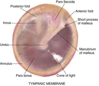
- Tympanic Membrane (Eardrum): A thin, translucent membrane that vibrates when struck by sound waves, forming the boundary between the outer and middle ear.
B. Middle Ear
The middle ear functions as a vital transmission chamber for sound vibrations. Its primary components include:
- Ossicles: Three tiny bones known commonly as the malleus (hammer), incus (anvil), and stapes (stirrup). These bones, collectively called ossicles, amplify and convey the vibratory signals from the eardrum to the inner ear.
- Eustachian Tube: A narrow passage connecting the middle ear to the nasopharynx, responsible for equalizing air pressure on both sides of the tympanic membrane, thereby ensuring optimal function.
- Tympanic Cavity: An air-filled space that houses the ossicles, essential for the proper conduction of vibrations.
C. Inner Ear
The inner ear is the most intricate part of the auditory system and is also known as the labyrinth. It comprises:
- Cochlea: A spiral-shaped, fluid-filled structure responsible for converting mechanical vibrations into electrical impulses that the brain interprets as sound.
- Vestibular System: It includes the semicircular canals, the vestibule, and the otolith organs, and plays a critical role in maintaining balance and spatial orientation.
Functions of Each Part of the Ear
- Outer Ear: Works as a funnel to capture and direct sound waves toward the eardrum.
- Middle Ear: Acts as a mechanical amplifier by transferring and amplifying vibrations via the ossicles while maintaining pressure equilibrium through the Eustachian tube.
- Inner Ear: Converts the mechanical vibrations originating in the middle ear into neural signals (via the cochlea) and manages balance through the vestibular apparatus.
How Sound is Processed and Transmitted
The transformation of sound into recognizable signals involves a remarkable and well-coordinated sequence of events. The process can be described as follows:
- Sound Collection: Sound waves are first gathered by the pinna and funneled through the external auditory canal.
- Vibration Initiation: As sound waves reach the tympanic membrane, they cause it to forcibly vibrate. This marked displacement is the initial mechanical response to sound.
- Amplification through Ossicles: The vibrations are transmitted to the ossicles in the middle ear. The ossicles act collectively as a lever system, amplifying the energy of the sound waves before they enter the inner ear.
- Transmission to the Inner Ear: The stapes, being the final link in the ossicle chain, pushes against the oval window, introducing the vibratory force into the fluid-filled cochlea.
- Conversion to Electrical Signals: Inside the cochlea, the movement of the perilymph and endolymph (fluid compartments) stimulates hair cells. These specialized receptor cells convert the mechanical energy into electrical impulses.
- Neural Transmission: The generated electrical signals travel along the auditory (cochlear) nerve to be processed by the brain, culminating in the perception of sound.
2. Assessment of Ear Health
Essential Components of Ear Health Evaluation
The clinical assessment of ear health is fundamental in recognizing and addressing auditory and balance disorders. This process is comprised of both thorough history taking and a detailed physical examination. Healthcare providers rely on a systematic approach to evaluate the ear, which includes examining both external structures and interior components using specialized tools.
A. History Taking
During history taking, clinicians explore the patient’s ear-related symptoms by asking targeted questions. The following areas are emphasized:
- Symptom Inquiry: Patients are queried about sensations such as ear pain (otalgia), hearing loss, tinnitus (persistent ringing), and vertigo (dizziness or imbalance).
- Onset and Duration: Understanding when symptoms began, their duration, and any associated changes (e.g., gradual or sudden onset) can provide clues about the underlying issue.
- Past Medical History: Questions about prior ear infections, trauma, exposure to loud noises, or previous surgeries help in formulating a differential diagnosis.
- Lifestyle and Occupational Factors: Factors such as noise exposure at work or during recreational activities are also assessed.
B. Physical Examination
The physical examination is a critical step in evaluating ear health, using both visual inspection and manual palpation to identify potential anomalies. Key aspects include:
- Visual Inspection: The external ear is examined for structural abnormalities, inflammation, or deformities. Observation of the ear canal for signs of infection or foreign bodies is also performed.
- Otoscopy: A specialized instrument called an otoscope is utilized to visualize the ear canal and the tympanic membrane. The appearance of the eardrum is meticulously examined for normal color, translucency, and tension, along with any signs of redness, retraction, bulging, or perforation.
- Palpation: Manual examination of the surrounding tissues and lymph nodes may be conducted to detect signs of infection or inflammation.
C. Common Signs and Symptoms to Observe
There are several key clinical indicators that warrant further investigation. These include:
- Ear Pain or Fullness: Pain may signify infection, trauma, or eustachian tube dysfunction.
- Hearing Loss: This symptom may be conductive (related to mechanical issues within the outer or middle ear) or sensorineural (linked to inner ear or nerve damage).
- Tinnitus: Persistent ringing or buzzing sounds can often be a symptom associated with ear damage or exposure to loud noises.
- Ear Discharge: Any exudate or leakage may indicate an infection or perforation of the eardrum.
- Dizziness/Vertigo: These symptoms are typically associated with disturbances in the vestibular system located in the inner ear.
D. Tools and Techniques for Ear Examination
The appropriate selection and application of diagnostic tools ensure an accurate assessment of ear health. Several instruments and tests are routinely used:
- Otoscope: This handheld device provides direct visualization of the ear canal and tympanic membrane, making it indispensable for identifying visible abnormalities.
- Tuning Fork Tests: Methods such as the Weber and Rinne tests help differentiate between conductive and sensorineural hearing loss by comparing bone conduction and air conduction thresholds.
- Tyampanometry: In some cases, tympanometry is used to measure the mobility of the tympanic membrane and middle ear pressure, providing insights into the mechanical function of the ear.
- Audiometry: This hearing test quantitatively evaluates hearing sensitivity over a range of frequencies, assisting in pinpointing the nature and extent of hearing loss.
3. Integrated Overview: Anatomy, Function, and Health Assessment
The following table consolidates key aspects of the ear’s structure, function, and examination techniques into one comprehensive view:
| Component | Key Structures | Primary Functions | Assessment Techniques |
|---|---|---|---|
| Outer Ear | Pinna, Ear Canal, Tympanic Membrane | Collects and directs sound waves; initiates vibration | Visual inspection, Otoscopy |
| Middle Ear | Ossicles (Malleus, Incus, Stapes), Eustachian Tube | Amplifies vibrations; transmits vibrations to inner ear; equalizes pressure | Otoscopy, Tuning Fork Tests, Tympanometry |
| Inner Ear | Cochlea, Vestibular System (Semicircular Canals, Otolith Organs) | Converts sound to electrical signals; maintains balance | Audiometry; specialized vestibular tests |
This table summarizes the integrated approach necessary for understanding ear anatomy, function, and clinical assessment. It highlights how each component is interdependent, forming a cohesive system that facilitates both auditory perception and balance.
References
-
Ear: Anatomy, Facts & Function - Cleveland Clinic
- Ear anatomy: Parts and functions - Kenhub
- How the Ear Works | Johns Hopkins Medicine
-
Ear Examination - StatPearls - NCBI Bookshelf
- Assessment of Ears: Video, Causes, & Meaning | Osmosis
Recommended Searches
Last updated March 27, 2025