
Unveiling Tomotherapy: How Precision Radiation Redefines Cancer Treatment
Discover the innovative technology that combines CT imaging with targeted radiation to combat cancer with remarkable accuracy.
Tomotherapy represents a significant advancement in radiation oncology, offering a sophisticated method for treating cancer. It ingeniously combines the detailed imaging capabilities of Computed Tomography (CT) with the precise radiation delivery of Intensity-Modulated Radiation Therapy (IMRT). This integrated approach allows clinicians to target tumors with exceptional accuracy while minimizing exposure to surrounding healthy tissues. Understanding how Tomotherapy works reveals its potential to transform cancer care for many patients.
Key Highlights of Tomotherapy
- Integrated Imaging & Therapy: Tomotherapy uniquely merges daily CT scanning with intensity-modulated radiation therapy (IMRT) in a single, streamlined device, enabling unparalleled treatment precision.
- Helical Delivery System: Radiation is administered in a continuous 360-degree spiral (helical) pattern, meticulously conforming the dose to the complex three-dimensional shape of the tumor while diligently sparing adjacent healthy tissue.
- Adaptive Treatment Capabilities: Daily on-board imaging allows oncologists to monitor changes in the tumor and patient anatomy, empowering them to adjust treatment plans in real-time for optimal targeting throughout the course of therapy.
Understanding Tomotherapy: A Synthesis of Precision and Imaging
At its core, Tomotherapy is a highly advanced form of image-guided radiation therapy (IGRT). Its unique architecture integrates two powerful technologies to deliver exceptionally precise treatments.
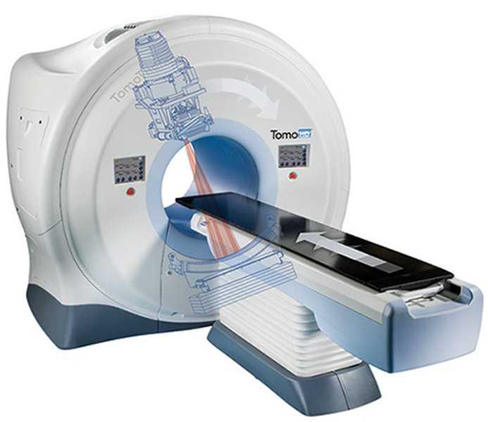
A TomoTherapy machine, integrating CT scanning and radiation delivery capabilities within a single unit.
Core Technologies: The Dual Power of Tomotherapy
Intensity-Modulated Radiation Therapy (IMRT)
IMRT is a sophisticated radiation delivery technique that allows the intensity of the radiation beam to be varied (modulated) across the treatment area. Tomotherapy employs a binary multileaf collimator (MLC) – a device with many small, computer-controlled "leaves" or "vanes" – that rapidly open and close to shape the radiation beam. This enables the creation of thousands of tiny "beamlets," each with its own intensity. By combining these beamlets from multiple angles as the gantry rotates, Tomotherapy can sculpt the high-dose radiation region to precisely match the three-dimensional contours of the tumor, even if it's irregularly shaped or wrapped around sensitive structures.
Integrated Computed Tomography (CT) Scanning
A key feature of Tomotherapy is its built-in CT scanner, typically using Mega-Voltage CT (MVCT). Before each treatment session, a 3D CT scan of the patient in the treatment position is acquired. This daily imaging, sometimes referred to as "Tomo-Graphy," provides up-to-date information on the tumor's exact size, shape, and location, as well as the position of nearby healthy organs. This is crucial because tumors can shrink or shift, and patient positioning can vary slightly from day to day. The system's software compares these daily images to the original planning CT scan, allowing for precise adjustments to ensure the radiation is delivered accurately every time.
The Tomotherapy Treatment Journey: Step-by-Step
The Tomotherapy process is a carefully orchestrated sequence of steps, ensuring that each patient receives a personalized and highly accurate treatment.
1. Meticulous Treatment Planning
The journey begins with a comprehensive treatment planning phase. The radiation oncology team, including radiation oncologists, medical physicists, and dosimetrists, uses high-resolution 3D images (often from a dedicated CT simulator, sometimes combined with MRI or PET scans) to precisely define the target tumor volume(s) and nearby critical organs and healthy tissues (Organs at Risk - OARs). Specialized software is then used to design an optimal IMRT plan. This involves determining the appropriate radiation dose for the tumor while setting strict dose limits for the OARs. The software calculates the complex pattern of beam intensities and angles required to achieve this highly conformal dose distribution from the Tomotherapy machine's helical delivery.
2. Patient Preparation and Precise Positioning
On the day of treatment, the patient is carefully positioned on the Tomotherapy machine's treatment table, typically lying on their back. To ensure reproducibility and minimize movement during treatment, custom immobilization devices (such as molds, masks, or cushions) may be used. Tiny, precise skin markings or tattoos, aligned with laser guides in the treatment room, help the radiation therapists position the patient accurately according to the treatment plan.
3. Daily Imaging and Verification: The Key to Accuracy
Once the patient is positioned, the integrated MVCT scanner acquires a 3D image of the treatment area. This scan usually takes only a few minutes. The system's software then automatically or with therapist guidance registers (aligns) this daily image with the reference images from the treatment plan. This comparison allows the team to verify the tumor's current position and make any necessary fine-tune adjustments to the patient's position on the table or, in some advanced adaptive radiotherapy protocols, to the radiation plan itself. This daily image-guidance is fundamental to Tomotherapy's precision, correcting for interfractional deviations caused by internal organ motion, tumor changes, or minor setup variations.
4. Helical Radiation Delivery: Targeting with Precision
With the tumor's location confirmed and adjustments made, the radiation delivery begins. The Tomotherapy machine features a linear accelerator (linac) mounted on a rotating ring gantry. As the gantry rotates 360 degrees around the patient, the treatment table moves slowly and precisely through the bore of the machine. This combination of gantry rotation and couch movement results in a helical (spiral) delivery pattern of radiation. During this process, the binary multileaf collimator continuously modulates the intensity of the thin, fan-shaped radiation beam, delivering thousands of optimized beamlets from a multitude of angles. This "slice-by-slice" or continuous rotational delivery ensures the radiation dose conforms tightly to the target volume.
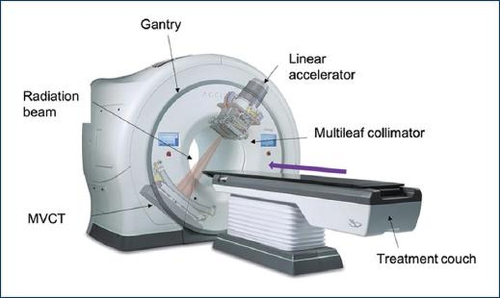
Diagram illustrating the helical (spiral) radiation delivery pattern characteristic of Tomotherapy, enabling 360-degree targeting.
5. Continuous Monitoring and Adaptation
Throughout the treatment session, the system is monitored by qualified radiation therapists. The daily imaging and precise delivery mechanisms inherently allow for a high degree of adaptability. Over the course of multiple treatment sessions (fractions), the accumulated data from daily imaging can inform oncologists about tumor response or anatomical changes, potentially leading to more significant plan adaptations if necessary, further personalizing the treatment.
Visualizing Tomotherapy's Key Attributes
To better understand Tomotherapy's capabilities in relation to other radiation modalities, the following chart provides a comparative assessment. It highlights attributes where Tomotherapy often demonstrates significant advantages. The scores are illustrative, representing general tendencies rather than absolute measures, and actual performance can vary based on specific clinical scenarios and equipment.
This chart visually compares Tomotherapy against conventional 3D conformal radiation therapy (3D-CRT) and standard non-helical IMRT across key performance indicators. Tomotherapy typically excels in precision targeting, sparing healthy tissues, adaptability due to its integrated daily imaging, and suitability for complex tumors, often leading to a more favorable side effect profile.
Mapping the Tomotherapy Workflow
The Tomotherapy process involves a series of interconnected stages, from initial assessment and planning through to the final delivery of radiation. This mindmap provides a visual overview of this comprehensive workflow.
This mindmap illustrates the sequential and integrated steps of the Tomotherapy process, emphasizing the cyclical nature of daily imaging and precise delivery that defines this advanced radiotherapy technique.
Advantages of the Tomotherapy Approach
Tomotherapy offers several distinct advantages over some conventional radiation therapy techniques, stemming largely from its integrated design and precision capabilities:
- Unmatched Precision and Accuracy: Daily CT imaging allows for highly accurate tumor targeting and adaptation to anatomical changes.
- Superior Sparing of Healthy Tissues: The IMRT and helical delivery enable the radiation dose to conform very tightly to the tumor, minimizing exposure to surrounding healthy organs and tissues. This can lead to fewer and less severe side effects.
- Adaptive Treatment Capabilities: The ability to adjust the treatment plan based on daily imaging ensures that the radiation remains targeted correctly, even if the tumor shrinks or shifts.
- Effective for Complex and Hard-to-Reach Tumors: Tomotherapy is particularly well-suited for treating tumors that are irregularly shaped, located near critical organs (e.g., head and neck, pancreas, spinal cord), or when multiple tumors need to be treated simultaneously.
- Treatment of Large or Elongated Tumors: The helical delivery mechanism allows for the treatment of long tumor volumes in a single, continuous pass, which can be advantageous for certain cancers.
- Potential for Dose Escalation: The precision of Tomotherapy may allow oncologists to safely deliver higher, more effective doses of radiation to the tumor.
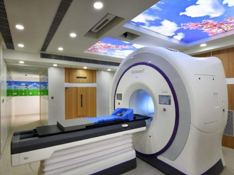
A modern Tomotherapy system, such as the Radixact X9, exemplifies the advanced technology used in precise cancer treatment.
The Patient Experience During Tomotherapy
What to Expect During a Session
For the patient, undergoing a Tomotherapy session is generally a straightforward and non-invasive experience. Each session typically lasts about 15 to 30 minutes, though this can vary. Much of this time is dedicated to patient positioning and the pre-treatment imaging verification; the actual radiation delivery may only take a portion of the total session time.
Patients lie on the treatment table, which moves smoothly through the ring-shaped gantry of the machine. The procedure itself is painless; patients do not feel the radiation as it is being delivered. They will hear the sounds of the machine operating, which is normal. It is important for the patient to remain as still as possible during imaging and treatment to ensure accuracy. The radiation therapists monitor the patient from a nearby control room via video and audio communication and can pause treatment if needed.
Potential Side Effects: What to Expect
While Tomotherapy's precision aims to minimize radiation exposure to healthy tissues, some side effects can still occur. These depend heavily on the area of the body being treated, the total radiation dose, and the individual patient's health. Most side effects are temporary and tend to resolve after the treatment course is completed.
- General Side Effects: Fatigue is a common side effect of radiation therapy. Skin reactions in the treated area, such as redness, dryness, or irritation (similar to a sunburn), may also occur.
- Localized Side Effects: Depending on the treatment site, specific side effects might include:
- Head and Neck: Dry mouth, taste changes, sore throat, difficulty swallowing, or localized hair loss.
- Chest: Cough, shortness of breath, or soreness when swallowing.
- Abdomen/Pelvis: Nausea, vomiting, diarrhea, or bladder irritation.
It is important for patients to discuss potential side effects with their oncology team, who can provide strategies for managing them. The precise targeting of Tomotherapy is designed to reduce the incidence and severity of these effects compared to less conformal radiation techniques.
Tomotherapy vs. Traditional Radiation: Key Differentiators
The following table provides a comparative overview of Tomotherapy and more traditional forms of external beam radiation therapy (EBRT), highlighting key differences in their technology and capabilities.
| Feature | Tomotherapy | Traditional External Beam Radiation Therapy (e.g., 3D-CRT) |
|---|---|---|
| Imaging Integration | Daily, on-board 3D CT (MVCT) imaging integrated into each treatment session for precise IGRT. | Less frequent imaging (e.g., weekly 2D port films or less frequent Cone-Beam CT), less integrated. |
| Radiation Delivery Method | Helical (spiral) delivery with 360-degree gantry rotation and continuous couch movement. | Typically uses static radiation fields from a few fixed angles or simple conformal arcs. |
| Beam Modulation | Highly sophisticated Intensity-Modulated Radiation Therapy (IMRT) using a binary multileaf collimator. | Limited or no intensity modulation (in basic 3D-CRT); IMRT can be done on conventional linacs but often with fewer beam angles than Tomotherapy. |
| Precision & Conformity | Extremely high, dose conforms very closely to complex tumor shapes. | Moderate conformity, may be less able to spare intricately located Organs at Risk (OARs). |
| Healthy Tissue Sparing | Superior, due to highly precise targeting, steep dose gradients, and comprehensive modulation. | Less effective at sparing healthy tissue, potentially leading to higher doses to surrounding structures. |
| Adaptability to Changes | High capacity for daily adjustments based on imaging; supports adaptive radiotherapy strategies. | Low; treatment plans are typically fixed for longer periods, with limited daily adaptation. |
| Suitability for Complex Tumors | Excellent, especially for tumors near critical organs, irregularly shaped tumors, or multiple targets. | Limited for very complex tumor shapes or locations requiring intricate OAR avoidance. |
| Treatment Verification | Volumetric 3D imaging (MVCT) performed daily before radiation delivery. | Often 2D planar imaging (kV or MV X-rays) or less frequent 3D Cone-Beam CT. |
See Tomotherapy in Action: Understanding the Process
This video provides a visual and narrated overview of the Tomotherapy process, explaining how it works and highlighting its benefits for cancer patients. It helps to conceptualize the sophisticated integration of imaging and treatment delivery that defines this advanced radiotherapy technique, offering insights into the patient experience and the technology's precision.
Frequently Asked Questions (FAQ)
Recommended Further Exploration
To deepen your understanding of Tomotherapy and related topics, consider exploring these questions:
- What are the specific advantages of Tomotherapy for prostate cancer treatment?
- How does the cost of Tomotherapy compare to other advanced radiation therapy options like CyberKnife or Proton Therapy?
- What are the latest advancements in Tomotherapy technology and adaptive radiotherapy?
- Are there any specific patient eligibility criteria or contraindications for Tomotherapy treatment?
References
Last updated May 14, 2025