
Unlocking the Mystery of Numerical Aperture Light Cones
Dive deep into how light collection and resolution hinge on NA and angular apertures.
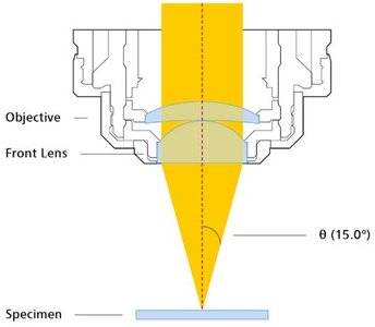
Highlights
- Definition and Formula: Numerical Aperture (NA) is defined as \(NA = n \cdot \sin(\theta)\), linking the refractive index and half-angular aperture.
- Light Cone Representation: The concept of light cones illustrates the range of angles captured by the objective, influencing resolution and brightness.
- Practical Applications: Understanding NA is crucial for selecting optimal objectives, whether using dry or immersion mediums, to enhance imaging quality.
Understanding Numerical Aperture and Light Cones
Definition and Basic Concept
The Numerical Aperture (NA) is a fundamental parameter in microscopy that quantifies the light-gathering ability of a microscope objective. Its mathematical definition is expressed by the formula:
\( \displaystyle NA = n \cdot \sin(\theta) \)
where:
- \( n \) is the refractive index of the medium between the specimen and the objective lens.
- \( \theta \) is half of the maximum angular aperture of the light cone that can be collected by the objective.
This formula encapsulates how both the refractive properties of the medium and the physical configuration of the lens contribute to the overall resolving power of the microscope.
The Role of Light Cones
In microscopy, the term "light cone" is used to describe the conical volume of light that can be collected or transmitted by the microscope objective. A higher numerical aperture corresponds to wider light cones, meaning that the objective can capture more oblique light rays. This is crucial because the ability to collect these rays directly correlates with the quality of image resolution.
As the numerical aperture increases, the objective’s light cone widens, allowing it to gather light in a wider range of angles. This increased capacity has two significant effects:
- Enhanced Resolution: More light, including high-angle rays, contributes to a finer detail in the captured image, allowing the microscope to resolve smaller structural details.
- Improved Brightness: A wider aperture collects more light overall, thereby enhancing the intensity and brightness of the resulting image.
Influence of the Imaging Medium
The imaging medium plays a critical role in determining the numerical aperture. Typically, with air as the medium (having a refractive index approximately equal to 1), there is an upper limit to the achievable NA (roughly 1.0). However, by using alternative immersion media such as water, glycerol, or oil, with refractive indices around 1.33, 1.47, and 1.51 respectively, one can achieve higher NA values. This enhancement allows microscopes to capture finer details, because:
- Immersion objectives provide a higher refractive index, directly boosting the value of \( n \) in our formula.
- Higher NA values result in a larger numerical aperture light cone, capturing more light and therefore raising the image resolution.
The proper selection of the imaging medium is paramount, especially in advanced microscopy techniques such as fluorescence and confocal microscopy.
Angular Aperture and Its Impact
The angular aperture is defined by the maximum angle \( \theta \) that the objective lens can accommodate from the specimen, often visualized as half of the light cone’s full angular extent. The relationship between angular aperture and numerical aperture is given by the sine function in the formula \( NA = n \cdot \sin(\theta) \).
A larger angular aperture means that the objective is capable of gathering light from a broader range of directions, thus collecting more diffracted rays. This greater collection directly improves image fidelity:
- Resolution: With a larger set of incoming angles, the ability to resolve closely spaced points enhances significantly.
- Light Collection: More diffused and oblique rays contribute to overall brightness and contrast of the image.
Practical Applications in Microscopy
Selection of Objectives
When choosing microscope objectives for a given application, one must consider not only the magnification but also the numerical aperture:
- Dry Objectives: Typically have lower maximum NA values (around 0.95) due to the air medium used between the specimen and the objective lens.
- Immersion Objectives: Use fluids such as oil or water, which result in higher NA values (up to 1.4 or higher). These are essential for high-resolution tasks, such as in cellular or sub-cellular imaging.
The careful balance between resolution and working distance is critical in applications like fluorescence imaging, where not only is detail important, but also the intensity and clarity of emitted light.
Interactive Learning Tools
To deepen one’s understanding of numerical aperture and its effect on light cones, interactive tutorials, like the one provided by MicroscopyU, offer valuable visualization tools. They allow users to dynamically adjust the angular aperture and observe changes in the light cone geometry. This hands-on experience helps connect theoretical formulas with real-world optical behavior, enhancing comprehension and application in research settings.
Comprehensive Comparison: Key Parameters in Light Cone Analysis
Below is a table summarizing the main parameters related to numerical aperture and how they influence the light cone in microscopy:
| Parameter | Description | Impact on Imaging |
|---|---|---|
| Numerical Aperture (NA) | \( NA = n \cdot \sin(\theta) \); quantifies the light-gathering capability. | Higher NA leads to improved resolution and brightness. |
| Refractive Index (n) | Represents the light refraction capabilities of the imaging medium (air, water, oil, etc.). | A higher refractive index increases NA, enabling collection of more light. |
| Angular Aperture (\( \theta \)) | Half of the total angle of the maximum light cone that can be collected. | Larger angles result in a wider light cone, enhancing resolution through increased light collection. |
| Objective Type | Dry vs. Immersion objectives, where immersion offers higher NA values. | Immersion objectives are preferred for high-resolution work due to their ability to use high-index media. |
Further In-Depth Discussion
Technical Insights
The relationship between light cone geometry and NA is central to optimizing microscope performance. When designing a microscope system or selecting components, understanding this interplay can lead to better instrument configurations for specific research applications. The broader the light cone, the more light the system collects. However, this also necessitates compromises in terms of depth-of-field, which is another important parameter in microscopy.
One must balance the desire for high resolution (high NA) with the practical limits imposed by working distances and the inherent trade-offs between resolution, brightness, and depth-of-field. For instance, while an oil immersion objective may offer exceptionally high NA and resolution, its extremely short working distance might limit its versatility in certain experimental setups.
Real-World Applications
Numerical aperture light cones are more than just theoretical constructs; they have direct implications in numerous scientific fields. In biological research, for example, reagents used in fluorescence microscopy require both high sensitivity and resolution. An objective with a high numerical aperture can capture faint fluorescent signals while resolving intricate details, helping researchers visualize internal cellular structures such as organelles or protein complexes.
Similarly, in materials science, the measurement of microstructures and fine-grained features relies heavily on the capabilities imparted by a high NA. Whether studying composite materials or semiconductors, the precision offered by advanced optical instruments underpins breakthroughs in understanding material properties at the microscale.
The systematic study of light cone behavior also informs the development of new imaging techniques that push the boundaries of resolution. Innovations in super-resolution microscopy, for example, build upon fundamental principles of NA and light collection efficiency to achieve imaging well beyond traditional diffraction limits.
References
- Numerical Aperture Light Cones | Nikon's MicroscopyU
- Numerical Aperture | Nikon's MicroscopyU
- Numerical Aperture and Light Cone Geometry - ZEISS
- Collecting Light: The Importance of Numerical Aperture in Microscopy - Leica Microsystems
- Numerical Aperture - Wikipedia
Recommended Related Queries
Last updated March 27, 2025