
Prostate Biopsy Unveiled: What to Expect from the Procedure and Your Results
A comprehensive guide to understanding prostate biopsies, from initial indications to navigating your options post-diagnosis.
A prostate biopsy is a crucial medical procedure for diagnosing prostate cancer. If your doctor has recommended one, or if you're seeking to understand this test better, this guide offers detailed insights into what it involves, the various types, preparation, recovery, and, importantly, the pathways available based on your test results. Understanding this process can empower you to make informed decisions about your health.
Essential Insights: Key Takeaways
- Definitive Diagnosis: A prostate biopsy is the primary method to definitively diagnose prostate cancer by examining tissue samples under a microscope.
- Multiple Approaches: Biopsies can be performed via different routes, including transrectal (most common), transperineal (lower infection risk), and MRI-guided techniques, each with specific advantages.
- Result-Driven Options: Biopsy results, including the Gleason score if cancer is found, are critical in determining the next steps, which can range from active surveillance for low-risk cancers to various treatment modalities for more aggressive forms.
Understanding the Prostate Gland and Biopsy Purpose
The prostate is a small gland found only in males, situated just below the bladder and in front of the rectum, surrounding the urethra (the tube that carries urine from the bladder). Its main function is to produce some of the fluid that makes up semen.
A prostate biopsy is recommended when initial tests suggest a potential problem with the prostate that requires closer examination. The primary goal is to detect the presence of cancerous cells. It is considered the gold standard for confirming a prostate cancer diagnosis.
When is a Prostate Biopsy Recommended?
Your healthcare provider may suggest a prostate biopsy if certain indicators point towards a risk of prostate cancer. These typically include:
- Elevated Prostate-Specific Antigen (PSA) Levels: PSA is a protein produced by the prostate gland. While elevated levels can be due to various conditions (like benign prostatic hyperplasia or prostatitis), a persistently high or rapidly rising PSA level is a common reason for a biopsy.
- Abnormal Digital Rectal Exam (DRE): During a DRE, a doctor inserts a gloved, lubricated finger into the rectum to feel the prostate for any lumps, hard areas, or other irregularities.
- Suspicious Imaging Results: If an MRI scan of the prostate reveals areas that look suspicious for cancer, a targeted biopsy of those areas might be recommended.
- Previous Biopsy Findings: If a prior biopsy showed abnormal cells that weren't cancerous (like high-grade Prostatic Intraepithelial Neoplasia or Atypical Small Acinar Proliferation), a repeat biopsy might be necessary.
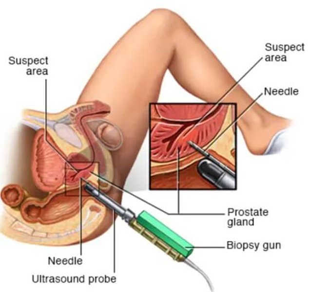
Illustration depicting a prostate biopsy procedure.
Types of Prostate Biopsy Procedures
There are several methods for performing a prostate biopsy, often utilizing imaging guidance to ensure accuracy.
Transrectal Ultrasound (TRUS)-Guided Biopsy
The Common Standard
This is historically the most common technique. An ultrasound probe is inserted into the rectum to create images of the prostate. A thin, spring-loaded needle is then passed through the rectal wall into the prostate to collect tissue samples (cores). Typically, 12 to 15 or more samples are taken from different regions of the prostate. Local anesthesia is used to numb the area.
Transperineal Biopsy
A Safer Alternative
In this method, the biopsy needle is inserted through the perineum (the skin between the anus and the scrotum). This approach is gaining popularity because it generally has a significantly lower risk of infection compared to the transrectal route, as the needle does not pass through the rectal wall. It can be performed under local or general anesthesia and may be guided by ultrasound or MRI. Transperineal biopsies can be:
- Targeted: Samples are taken from specific areas that appeared suspicious on prior MRI scans.
- Template: A grid is placed over the perineal skin, and samples are taken systematically.
MRI-Guided or MRI/Ultrasound Fusion Biopsy
Precision Targeting
This advanced technique uses Magnetic Resonance Imaging (MRI) to identify suspicious areas within the prostate. During the biopsy, these MRI images can be fused with live ultrasound images (MRI/Ultrasound Fusion Biopsy) or the biopsy can be performed directly within an MRI scanner (in-bore MRI-guided biopsy). This allows for highly targeted sampling of suspicious lesions, potentially improving the detection rate of clinically significant cancers and reducing the number of cores needed.
Transurethral Biopsy
Less Common Approach
This method is used less frequently. A cystoscope (a thin tube with a camera) is inserted through the urethra to reach the prostate. Tissue samples are then taken through the scope. This procedure is typically done with medication to make the patient sleepy and prevent pain.
Preparing for Your Prostate Biopsy
Proper preparation is key to a smooth procedure and minimizing risks.
- Antibiotics: You will likely be prescribed antibiotics to take before and after the biopsy to prevent infection. It's crucial to complete the entire course as directed.
- Medication Review: Inform your doctor about all medications you take, especially blood thinners (like aspirin, warfarin, clopidogrel), as you may need to stop them temporarily before the procedure.
- Dietary Instructions: You might be asked to use an enema or follow specific dietary restrictions before the biopsy, particularly for transrectal procedures. If sedation or general anesthesia is planned, you'll need to fast for several hours beforehand.
- Arrange Transportation: If you receive sedation or general anesthesia, you will not be able to drive yourself home, so arrange for someone to accompany you.
During the Prostate Biopsy Procedure
The procedure itself usually takes between 10 to 45 minutes, depending on the type and imaging guidance used.
- Positioning: You will typically lie on your side with your knees bent towards your chest, or sometimes on your back with your legs in stirrups.
- Anesthesia: Local anesthesia (an injection to numb the prostate area) is almost always given to minimize discomfort. Some procedures, especially transperineal or MRI-guided ones, may involve sedation or general anesthesia.
- Imaging Guidance: An ultrasound probe (for TRUS or transperineal) will be used to visualize the prostate. For MRI-guided procedures, you'll be positioned within or near an MRI scanner.
- Sample Collection: A spring-loaded instrument is used to quickly insert a thin needle and retrieve small, cylindrical cores of prostate tissue. You might hear a clicking or snapping sound with each sample. Multiple samples (often 12 or more) are taken from different parts of the prostate.
Comparing Biopsy Techniques: A Visual Overview
Different prostate biopsy techniques offer varying balances of accuracy, invasiveness, and potential side effects. The radar chart below provides a comparative look at some key aspects of common biopsy methods. This is a generalized comparison; individual experiences may vary. Values are scaled from 1 (less favorable) to 5 (more favorable) for each attribute, based on general medical understanding.
This chart illustrates that while TRUS biopsy is common, newer methods like transperineal and MRI-fusion biopsies may offer advantages in terms of lower infection risk and higher precision for detecting significant cancers.
Recovery and Post-Biopsy Care
After the biopsy, you'll be monitored for a short period before going home. Recovery is usually quick, but some side effects are common:
- Soreness: Mild pain or discomfort in the rectal or perineal area is normal and can be managed with over-the-counter pain relievers like acetaminophen. Avoid aspirin or ibuprofen unless cleared by your doctor, as they can increase bleeding.
- Blood in Urine (Hematuria): Pink or red-tinged urine is common for a few days.
- Blood in Semen (Hematospermia): Semen may appear reddish or rust-colored for several weeks or even a couple of months. This is normal and harmless.
- Blood in Stool: Light rectal bleeding may occur for a few days, especially after a transrectal biopsy.
- Activity Restrictions: Your doctor will likely advise you to avoid strenuous activities, heavy lifting, and sexual intercourse for a few days to a week to allow the area to heal and reduce irritation.
Contact your doctor immediately if you experience fever, chills, difficulty urinating, heavy or persistent bleeding, or worsening pain, as these could be signs of an infection or other complication.
Navigating the Prostate Biopsy Journey: A Mindmap Overview
The journey from initial concern to understanding biopsy results and subsequent options can be complex. This mindmap provides a visual summary of the key stages involved in the prostate biopsy process.
(Blood in urine/semen, etc.)"] id4c["Activity Restrictions"] id4d["When to Call Doctor"] id5["Biopsy Results Analysis"] id5a["Pathology Report"] id5b["Gleason Score / Grade Group"] id5c["Presence of Cancer"] id5d["Other Findings (PIN, ASAP)"] id6["Options Based on Results"] id6a["Negative for Cancer
(Monitoring)"] id6b["Abnormal Cells (Not Cancer)
(Further Tests/Repeat Biopsy)"] id6c["Cancer Detected"] id6c1["Active Surveillance"] id6c2["Surgery (Prostatectomy)"] id6c3["Radiation Therapy"] id6c4["Hormone Therapy"] id6c5["Other Treatments"]
This mindmap outlines the typical progression, highlighting the critical decision points and outcomes at each step of the prostate biopsy process.
Interpreting Your Biopsy Results and Subsequent Options
The tissue samples collected during the biopsy are sent to a pathologist, who examines them under a microscope. Results are usually available within a few days to a week. Your doctor will discuss these results with you and explain what they mean for your health.
Negative for Cancer
If no cancer cells are found, this is reassuring. However, biopsies can sometimes miss small cancers. Depending on your PSA levels, DRE findings, and other risk factors, your doctor may recommend:
- Continued monitoring with regular PSA tests and DREs.
- An MRI scan if not already done, to look for suspicious areas that might have been missed.
- A repeat biopsy at a later date, especially if PSA levels continue to rise or other concerns persist.
Abnormal Cells (Not Cancerous, but Potentially Pre-Cancerous)
Sometimes, the biopsy may reveal cells that are not cancerous but are not entirely normal. These include:
- Prostatic Intraepithelial Neoplasia (PIN):
- Low-grade PIN: Generally not a cause for concern, as many men have it.
- High-grade PIN: Considered a pre-cancerous condition. Finding high-grade PIN may increase the likelihood of finding cancer on a subsequent biopsy. Your doctor might recommend closer follow-up or a repeat biopsy sooner.
- Atypical Small Acinar Proliferation (ASAP): This finding means that abnormal cells were seen, but it's unclear if they are cancerous. ASAP significantly increases the chance of finding prostate cancer on a repeat biopsy, which is often recommended.
Prostate Cancer Detected
If cancer cells are found, the pathologist's report will include important information about the cancer, primarily its grade and sometimes its volume (how much cancer is in the samples).
Understanding the Gleason Score and Grade Group
The aggressiveness of prostate cancer is most commonly graded using the Gleason score. The pathologist identifies the two most common cancer cell patterns and assigns a grade from 1 to 5 to each (5 being the most abnormal). These two grades are added together to give a Gleason score (e.g., 3+4=7).
More recently, a simpler Grade Group system (from 1 to 5) has been adopted:
- Grade Group 1: Gleason score 6 or less (low-grade cancer, tends to grow slowly)
- Grade Group 2: Gleason score 3+4=7 (intermediate-grade, favorable)
- Grade Group 3: Gleason score 4+3=7 (intermediate-grade, unfavorable)
- Grade Group 4: Gleason score 8 (high-grade cancer)
- Grade Group 5: Gleason scores 9-10 (very high-grade cancer, likely to grow and spread quickly)
Along with your PSA level, DRE findings, and imaging results (like MRI), the Gleason score/Grade Group helps determine the cancer's stage (how far it has spread) and risk group (low, intermediate, or high risk of progression). This information is crucial for deciding on the best course of action.
Treatment Options Based on Cancer Diagnosis
If prostate cancer is diagnosed, various options are available. The choice depends on many factors, including the cancer's grade and stage, your age and overall health, potential side effects, and personal preferences.
Common approaches include:
| Result Category / Risk Group | Typical Gleason Score / Grade Group | Common Management & Treatment Options |
|---|---|---|
| Low-Risk Localized Cancer | Gleason 6 (Grade Group 1) |
|
| Intermediate-Risk Localized Cancer | Gleason 7 (Grade Group 2 or 3) |
|
| High-Risk Localized Cancer | Gleason 8-10 (Grade Group 4 or 5) |
|
| Locally Advanced or Metastatic Cancer (Spread beyond the prostate) | Varies, often higher grades |
|
This table provides general guidelines. Your specific treatment plan will be individualized by your healthcare team.
Further tests, such as CT scans, bone scans, or more advanced imaging like PSMA PET scans, may be done to determine if the cancer has spread outside the prostate, which also influences treatment choices.
Understanding Your Biopsy: A Deeper Dive
The following video provides a helpful overview of what patients should know about the prostate biopsy procedure, complementing the information discussed.
This video from the Urology Care Foundation features Dr. John Lynch discussing what patients should know about a prostate biopsy.
Alternatives to Immediate Biopsy or Tests Aiding Biopsy Decisions
While a biopsy is the only way to definitively diagnose prostate cancer, several tests can help assess risk and determine if a biopsy is necessary, potentially avoiding unnecessary procedures for some men:
- Multi-parametric MRI (mpMRI): This advanced imaging scan provides detailed pictures of the prostate and can identify suspicious areas. A normal mpMRI may sometimes lead to deferring a biopsy in men with slightly elevated PSA. If suspicious areas are seen, it can guide a targeted biopsy.
- Advanced Blood and Urine Tests: Newer biomarker tests (e.g., Prostate Health Index (PHI), 4Kscore, SelectMDx, ExoDx Prostate Test) can provide additional risk stratification beyond PSA alone, helping to decide who might benefit most from a biopsy.
These tools do not replace biopsies but aid in shared decision-making between you and your doctor.
Frequently Asked Questions (FAQ)
What is a prostate biopsy?
Is a prostate biopsy painful?
How long does it take to get prostate biopsy results?
What are the main risks of a prostate biopsy?
What is a Gleason score?
Conclusion
A prostate biopsy is a vital diagnostic tool in the detection and management of prostate conditions, particularly prostate cancer. Understanding the procedure, from preparation through recovery and interpretation of results, can help alleviate anxiety and empower you to participate actively in your healthcare decisions. The options available after a biopsy are tailored to the specific findings and your individual circumstances, always aiming for the best possible outcome. Open communication with your healthcare team is essential throughout this process.
Recommended Further Exploration
- What are the long-term side effects of different prostate cancer treatments?
- How does active surveillance for prostate cancer work and who is it suitable for?
- Can lifestyle changes impact prostate cancer risk or progression?
- What advancements are being made in prostate cancer diagnosis and treatment?
References
Last updated May 21, 2025