
Unlocking Gastric Secrets: How the Rat Stomach Illuminates Human Disease
Discover the intricate histological landscape of the rat stomach to inform our understanding of human gastric pathologies
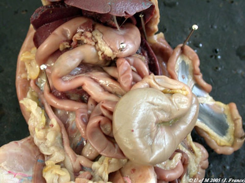
Key Highlights
- Distinct Functional Zones: The forestomach and glandular stomach exhibit unique histological profiles that are fundamental to understanding gastric pathology.
- Comparative Insights: Comprehensive histological analysis of the rat stomach provides an effective model for exploring gastric ulcers and metaplasia in humans.
- Research & Pathology Applications: Detailed examinations in both gross anatomy and cellular histology enhance our grasp of disease progression and treatment strategies.
Detailed Histological Characterization of the Rat Stomach
The rat stomach is a complex organ comprising distinct histological zones that include the non-glandular forestomach and the glandular stomach. The comparative examination of these regions reveals meaningful insights into gastric pathologies that parallel certain aspects of human stomach diseases. This analysis not only underscores structural similarities but also highlights unique features that empower scientific research using the rat model.
A Closer Look at Functional Zones
The Forestomach
The forestomach occupies the proximal region of the rat stomach and is characterized primarily by a non-glandular, stratified squamous epithelium. This keratinized protective lining serves as a preliminary storage and processing area for ingested food. The forestomach's robust epithelium is ideally structured to withstand mechanical abrasion resultant from rough, fibrous diets. However, this adaptation also predisposes the forestomach to specific pathologies, such as ulcerations under stress conditions (e.g., protein restriction or exposure to toxins).
Histologically, the forestomach does not participate in enzymatic digestion; instead, it functions as a reserve compartment where initial processing occurs before food passes into the glandular region. Its distinct histological profile, including a lack of specialized secretory cells, makes it an important area of study especially when exploring non-glandular pathologies and the onset of inflammation or metaplastic changes.
The Glandular Stomach
In contrast, the glandular stomach is subdivided into distinct regions—the fundus, corpus (body), and pylorus (antrum). This portion of the stomach is lined with simple columnar epithelium and is richly endowed with specialized secretory cells responsible for the production of digestive enzymes and acids. Prominent among these cells are:
- Parietal Cells: These cells secrete hydrochloric acid (HCl), facilitating the breakdown of ingested food and providing a hostile environment for ingested pathogens. They also produce intrinsic factor, essential for vitamin B12 absorption.
- Chief Cells: Responsible for the synthesis and secretion of pepsinogen, the zymogen precursor of the enzyme pepsin, these cells are fundamental to protein digestion.
- Mucous Neck Cells: These cells produce mucus that provides a protective barrier, safeguarding the mucosal lining from the corrosive effects of gastric acid.
The glandular stomach is pivotal in advanced digestive processing and is a key site for pathological changes such as atrophy, metaplasia, and even neoplastic transformations. Comparative studies indicate that alterations in this region in rats bear a resemblance to the progression of similar conditions in humans, making it an invaluable model for research.
Histological and Muscular Architecture
Muscular Organization
The muscularis externa of the rat stomach comprises three definitive layers:
- Longitudinal Muscle: This outer muscle layer provides the structural framework for the stomach, facilitating gross movement and mixing of stomach contents.
- Circular Muscle: Positioned beneath the longitudinal layer, it plays a critical role in the churning and peristaltic mixing of ingested substances.
- Internal Oblique (Sling) Muscle: Particularly prominent at the gastro-esophageal junction, this layer supports the transition between the esophagus and the stomach, with some extensions of striated muscle into the distal esophagus distinguishing it from its human counterpart.
This layered muscular arrangement is essential to efficient gastric motility and correlates with clinical observations in gastric motility disorders and ulcerations. Detailed histological studies further reveal the differential distribution of muscle fibers that can impact both the mechanical and secretory functions of the stomach.
Comparative Histopathology in Disease Models
The histological study of both the forestomach and glandular stomach helps delineate changes associated with various gastric pathologies. In experimental settings, researchers have documented instances of:
- Gastric Ulcers: Both regions, especially the glandular stomach, respond to injurious stimuli (e.g., chemicals and stress) through ulcer formation. The progression of ulceration in rats provides cues on inflammatory responses and tissue repair mechanisms relevant to human medicine.
- Metaplasia: Chronic exposure to irritants and sustained inflammatory states can induce metaplastic changes in the gastric mucosa. These cellular transformations in the rat model often mimic early stages of pre-cancerous transformations observed in human gastric tissues.
Given these parallels, the rat stomach emerges as an outstanding model for studying the morphological and functional evolution of gastric diseases. The detailed histological characterizations have facilitated a deeper understanding of cellular responses, glandular adaptations, and muscular remodeling under pathological conditions.
Visualizing the Data: Integrated Comparisons
Comprehensive Comparison Table
The table below summarizes the salient features of the rat stomach as compared to human gastric anatomy, focusing on crucial histological and functional aspects.
| Features | Rat Stomach | Human Stomach |
|---|---|---|
| General Structure | Divided into forestomach (non-glandular) and glandular regions | Primarily glandular with no forestomach |
| Epithelium | Forestomach: Stratified squamous keratinized; Glandular: Simple columnar | Simple columnar throughout the mucosal layer |
| Gastric Secretions | Fundic glands secrete pepsinogen and HCl; presence of mucous cells | Rich secretory cells including parietal, chief, and mucous cells |
| Muscular Layers | Three layers: longitudinal, circular, and oblique (sling) muscle | Similar trifold arrangement but with differences in extent of striated muscle at the esophageal junction |
| Pathological Susceptibility | Demonstrates ulcer formation and metaplastic changes under stress conditions | Exhibits ulcers, metaplasia, and atrophic gastritis under chronic stress |
Integrating Research Methodologies
Detailed histological evaluations are executed using techniques such as light and electron microscopy, which reveal the ultra-structural details of gastric mucosa, including the arrangement of microvilli, the integrity of cellular organelles, and the composition of intercellular junctions. These insights are critical in both baseline research and in the diagnosis of pathological states such as gastritis and neoplasia.
Studies leveraging the rat model have utilized various immunohistochemical staining techniques to map out the distribution of cellular markers such as Ki-67 for proliferative activity and cytokeratins for epithelial integrity. Such analyses enhance our understanding of disease progression and help identify potential therapeutic targets.
Radar Chart Analysis: Functional Contributions in the Rat Stomach
The interactive radar chart below provides a conceptual overview of different histological parameters of the rat stomach. The multiple datasets reflect opinionated analyses of key aspects such as cellular specialization, muscular structure, secretory functionality, and vulnerability to pathology. This visualization models the comparative strengths of each domain, helping bridge the gap between structure and function.
Supplementary Multimedia Resource
For an in-depth visualization of rat dissection which includes detailed exposure of the abdominal cavity and the gastrointestinal system, view the video below. It provides context for the anatomical positioning and relative structure of the stomach, enhancing your understanding of the material covered.
Frequently Asked Questions
What histological techniques are commonly used to study the rat stomach?
Researchers typically use a combination of light and electron microscopy to examine the cellular architecture of the rat stomach. Immunohistochemical staining is also widely used to detect cellular markers such as Ki-67, cytokeratins, and various digestive enzymes. These methods allow for detailed visualization of the histopathology associated with gastric ulcers, metaplasia, and other pathologies.
How do the functional zones of the rat stomach correlate with human gastric anatomy?
Although the rat stomach uniquely contains a forestomach – which is absent in humans – the glandular region in rats showcases many similarities to the human stomach. Both possess specialized cells responsible for acid and enzyme secretion, and both experience similar pathological changes such as ulceration and metaplasia in the glandular region. This comparative model enhances our understanding of human gastric diseases.
Why is the rat stomach a preferred model for studying gastric ulcers and metaplasia?
The rat stomach is favored due to its distinct yet comparable histological features when contrasted with the human stomach. The presence of both a non-glandular forestomach and a glandular region, along with similar responses to injurious stimuli, allows for the exploration of ulcerative processes and metaplastic changes which are relevant to understanding human gastric pathologies. Moreover, the reproducibility of these histological findings in rats furthers research into therapeutic strategies.
References
-
Surgical Anatomy of the Gastrointestinal Tract and Its Vasculature - PMC
- Anatomy, Histology, Ultrastructure, Stomach, Rat - Springer
- Rat Anatomy - Clarendon College
- Histology of the Stomach in Control Rats - ResearchGate
-
Organisation of the Musculature of the Rat Stomach - PMC
Recommended Further Queries
Last updated April 2, 2025