
Research Paper Article Format: Comparative Histology of Rat and Human Stomachs
A Step-by-Step Guide with Detailed Examples for a 3000-Word Scientific Article
Highlights
- Structured Format – Includes Title Page, Abstract, Introduction, Methods, Results, Discussion, Conclusion, and References.
- Detailed Examples – Each section is illustrated with practical guidelines and sample texts to aid in writing a comprehensive 3000-word paper.
- Comparative Emphasis – Focuses on highlighting key differences and similarities in the histology of the rat stomach versus the human stomach.
1. Title Page
Example Title Page
Title: Comparative Histological Analysis of the Rat and Human Stomach
Authors: [Your Name], [Collaborator’s Name]
Institutional Affiliation: Department of Anatomy, [Your University/Institution]
Date: [Month Day, Year]
Note: The title page is the first page of your paper and should clearly state the title along with the authors’ names, affiliations, and the submission date. Use a standard 12 pt Times New Roman font with 1-inch margins.
2. Abstract
Abstract Section with Example
Abstract: (150-250 words)
The objective of this study is to compare and contrast the histological architecture of the rat stomach with that of the human stomach. Using standard histological procedures, tissue samples were processed, sectioned, and stained with hematoxylin and eosin (H&E) as well as special stains such as Periodic Acid-Schiff (PAS) and Alcian Blue. The rat stomach is characterized by a distinct non-glandular forestomach region and a glandular portion, while the human stomach is uniformly glandular. All examined layers – mucosa, submucosa, muscularis externa, and serosa – were compared with an emphasis on glandular distribution, cellular composition, and mucosal architecture. The findings provide a detailed comparative insight that is essential for the accurate interpretation of data derived from rat models in gastrointestinal research. These histological insights have significant implications for the translation of experimental results from animal studies to human pathophysiology.
Note: The abstract should be concise and summarize the research question, methodology, key findings, and overall significance.
3. Introduction
Introduction with Background and Research Context
The stomach is a vital organ responsible for the initiation of digestion in mammals. Comparative histological studies between species, notably the rat and human, provide valuable insights that can enhance the interpretation of biomedical research. Rats are commonly used in experimental studies due to their ease of handling and genetic similarities to humans. However, inherent anatomical and histological differences must be carefully considered. This paper is centered on a comprehensive analysis of the histological structures of the rat stomach compared with the human stomach.
Historically, the rat model has played a crucial role in digestive system research. An important feature of the rat stomach is the presence of a non-glandular forestomach, which is typically absent in humans. In humans, the stomach is entirely glandular, with subdivisions into the cardia, fundus, body, and pylorus. This differentiation is not only morphological but also functional; the protective and secretory roles of the epithelial layers are critical for understanding species-specific digestive physiology.
This section outlines the rationale behind the comparative approach and addresses the following research questions:
- What are the key histological differences between the rat and human stomach?
- How do these differences affect the interpretation of rat models in gastrointestinal research?
- What implications do these findings have for translational research in human gastric diseases?
Additionally, a comprehensive review of prior literature suggests that while similarities exist in basic stomach architecture across species, notable differences in cellular lining, glandular structures, and muscular composition underscore the need for a tailored approach when extrapolating results.
4. Materials and Methods
Methodological Approach and Protocols
The Materials and Methods section details the experimental design, sample collection, preparation, and subsequent slide analysis for histological sections. Adhering to standard ethical guidelines is imperative.
Sample Collection: Rat stomach samples were collected from adult Sprague-Dawley rats under approved ethical guidelines. Human stomach tissue samples were obtained from consenting patients during routine endoscopic or biopsy procedures.
Tissue Preparation: All tissue samples were fixed in 10% neutral buffered formalin for 24 hours to preserve structural integrity. Following fixation, samples were processed through graded alcohols and embedded in paraffin. Sections were cut at a thickness of 5 μm.
Staining Procedures: Standard Hematoxylin and Eosin (H&E) staining was employed for general tissue morphology. To specifically highlight mucin components and cellular variations, stains like Periodic Acid-Schiff (PAS) and Alcian Blue were used.
Microscopic Examination: Slides were examined under a light microscope with varying magnifications to assess cellular morphology, gland distribution, and layer thickness. Digital image analysis software was used to quantify histological variations.
Note: Detailed protocols for sample processing and staining ensure the reproducibility of the experimental results and provide a foundation for comparing histological differences across species.
5. Results
Presentation of Histological Findings
The results section presents a systematic comparison of histological features observed in the rat and human stomach samples. The following table summarizes key histological characteristics:
| Feature | Rat Stomach | Human Stomach |
|---|---|---|
| Mucosal Layer | Thin in the non-glandular forestomach; thicker in glandular region | Uniformly thick glandular mucosa |
| Epithelial Lining | Stratified squamous epithelium in forestomach; simple columnar in glandular region | Simple columnar epithelium throughout |
| Glandular Structure | Localized distribution with a higher concentration of parietal and chief cells in the fundic region | Even distribution across the fundus and body |
| Muscularis Externa | Includes additional oblique muscle layer | Classical three-layer structure (longitudinal, circular, and oblique muscle layers) |
| Additional Observations | Presence of a limiting ridge dividing forestomach and glandular stomach | No distinct non-glandular region observed |
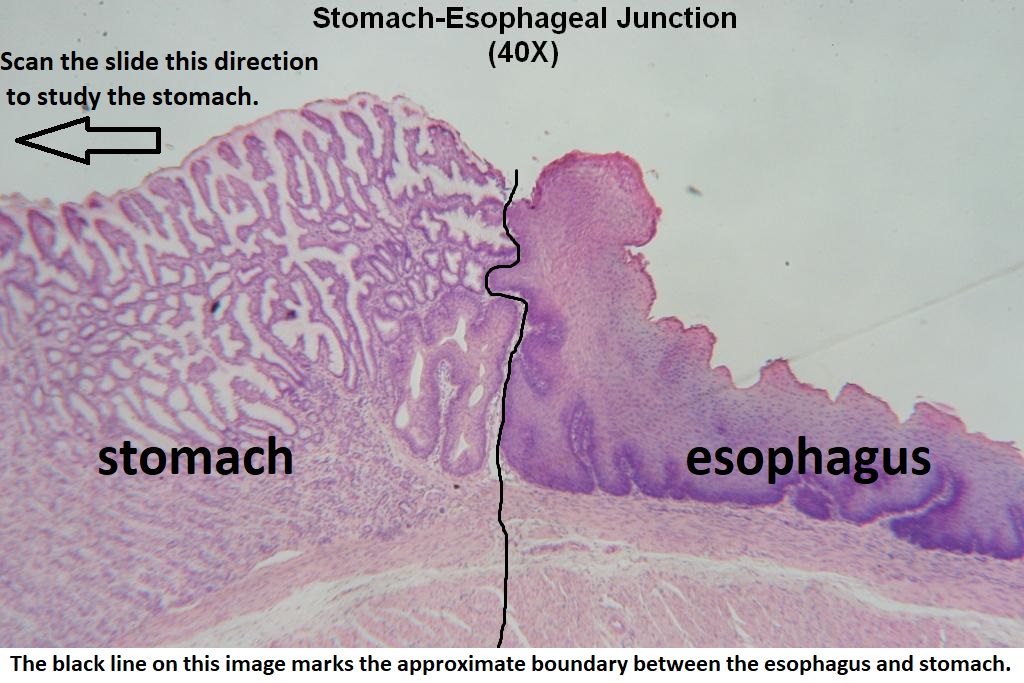
Figure Example: Light microscopy images further illustrate the differences in cellular architecture. For instance, the stratified squamous epithelium in the rat forestomach contrasts sharply with the homogeneous columnar epithelium of the human stomach tissue.
In the glandular regions, quantitative analysis indicated a statistically significant variation in mucosal thickness and gland density. These parameters were measured using image analysis software and confirmed through repeated sampling.
Additional observations include the comparative density of parietal cells and the distribution of chief cells. Such differences underscore the biological implications of utilizing rat models for research into human disease processes, as variations in digestive enzyme secretion and acid production were noted.
6. Discussion
Interpretation and Implications of Findings
The discussion section synthesizes the results and provides interpretations within the framework of comparative anatomy and physiological relevance. One of the most striking differences observed is the presence of a non-glandular forestomach in rats, which provides an adaptation for the initial processing of ingested food. This region, lined by stratified squamous epithelium, contrasts with the uniformly glandular human stomach, suggesting differences in function and digestive efficiency.
Further analysis reveals that while both species share a common layered stomach architecture consisting of the mucosa, submucosa, muscularis externa, and serosa, the distribution and density of key cellular components vary. In particular:
- The glandular portion of the rat stomach exhibits a higher localized density of parietal cells, especially in the fundic region, which may influence gastric acid secretion patterns.
- Similarly, the distribution of chief cells in the rat stomach appears more region-specific, potentially impacting the secretion of digestive enzymes compared to the more uniform distribution seen in human tissue.
- The additional oblique muscle layer present in the rat stomach could contribute to differences in mechanical digestion, an aspect that warrants further biomechanical studies.
The implications of these findings extend to experimental design and the extrapolation of data from rat models to human clinical research. For instance, the presence of a forestomach in rats – absent in humans – should be taken into account when studying drug absorption and the localized effects of ingested substances. This fundamental difference in gastrointestinal anatomy highlights the necessity for careful methodological cross-validation when interpreting preclinical data.
Moreover, our findings correlate with previous studies where species-specific variations in gastric histology were shown to affect results in pharmacological testing and disease modeling. The variations in mucosal thickness and glandular cell distribution imply that while rats serve as valuable models for human gastric studies, results must be contextualized within the framework of these inherent histological differences.
Moving forward, further research should focus on the functional implications of these histological differences. Studies incorporating immunohistochemical markers and molecular analyses could provide deeper insights into the regulatory mechanisms underpinning these variations. Additionally, future investigations could explore the translational relevance of these differences in the context of gastric pathologies and therapeutic interventions.
7. Conclusion
Summary of Key Comparative Insights
In conclusion, this comparative study of the rat and human stomach histology highlights several critical insights. The rat stomach exhibits a distinct dichotomy between a non-glandular forestomach and a glandular region, while the human stomach is entirely glandular across its anatomical regions. This study elucidates significant differences in mucosal thickness, glandular cell distribution, and muscular layers. Such variations not only impact digestive physiology but also have profound implications for translational research utilizing rat models.
The comprehensive analysis provided herein emphasizes the necessity of accounting for species-specific anatomical variations when interpreting experimental findings. The meticulous characterization of cytological features — including the distribution of parietal and chief cells — reinforces the importance of aligning histological data with clinical applications in human health research. Ultimately, while rat models remain invaluable for experimental purposes, a nuanced approach is required to bridge the gap between animal studies and human clinical outcomes.
The presented format serves as a detailed template for constructing a 3000-word research paper, ensuring that all essential sections are covered, and research findings are presented with clarity and scientific rigor. Researchers are encouraged to adopt and adapt this structured approach to further explore the implications of histological differences between species.
8. References
Citing the Literature
All references should adhere to a consistent citation style such as APA, MLA, or Chicago. Below is an example list of references that might be used in this research paper:
- Stomach Histology - Kenhub
- Research Paper Format - Scribbr
- Sample APA Papers - APA Style
- Gastric Histology - University of Leeds
- Comparative Anatomy in Rats - Wiley Online Library
Conclusion and Final Thoughts
Summary of the Research Paper Format
This guide presents a comprehensive and detailed format for writing a 3000-word research paper comparing the histology of the rat and human stomachs. It emphasizes a logical progression from the title page and abstract to the detailed experimental sections, culminating in an integrative conclusion. Each section is crafted to showcase both structure and specificity, ensuring that your paper is not only scientifically rigorous but also accessible to readers from diverse academic backgrounds.
By adhering to this format, researchers can effectively communicate their findings and contribute valuable insights into comparative gastrointestinal histology. The step-by-step examples provided in this guide are designed to ensure that essential elements are covered, offering a solid framework for the development of well-organized and coherent scientific manuscripts.
Recommended Related Queries
Deepen Your Research Exploration
Last updated February 22, 2025