
Surgical Case Report: Unilateral Inguinal Hernia and Cryptorchidism in a 3-Year-Old Chihuahua
A detailed surgical case report integrating clinical presentation, diagnostics, intervention, and follow-up care
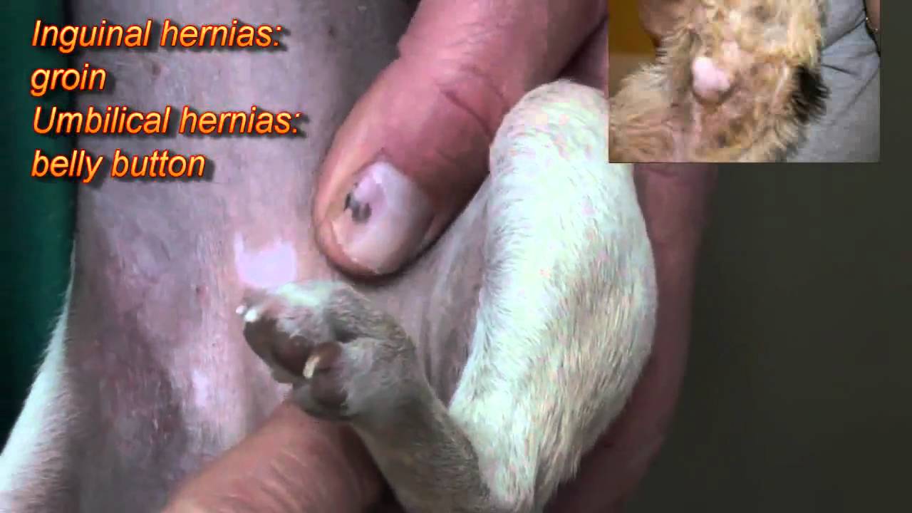
Key Highlights
- Comprehensive Clinical and Diagnostic Approach: Detailed physical examination, imaging evaluations, and relevant laboratory tests provided crucial insights for an accurate diagnosis.
- Surgical Techniques and Management: The case report outlines specific surgical maneuvers including herniorrhaphy, orchiectomy, and postoperative care planning.
- Postoperative Recovery and Follow-Up: Emphasis on post-surgery monitoring, pain management, and long-term follow-up ensures a favorable outcome.
Introduction
In veterinary medicine, surgical case reports provide significant insights into the management of complex conditions encountered in clinical practice. This report addresses a case involving a 3-year-old Chihuahua presenting with a unilateral inguinal hernia complicated by cryptorchidism. Notably, the Chihuahua's predisposition to both inguinal hernias and testicular maldescent underscores the importance of a thorough diagnostic and surgical intervention approach, ensuring the animal’s well-being and minimizing potential complications.
Clinical Presentation
Patient Overview
The patient in this report is a 3-year-old male Chihuahua presented with swelling in the right inguinal region. The owner had noted a palpable enlargement over several weeks, and the animal exhibited no overt signs of distress apart from localized discomfort. The patient’s physical profile further revealed the absence of the right testicle in the scrotum, with only the left testicle normally descended.
Historical and Physical Examination Data
Key observations during the initial clinical evaluation included:
- History: The dog had no significant previous medical issues, and the swelling was a recent development noticed by the owner.
- Physical Examination Findings: A palpable mass in the right inguinal canal consistent with an inguinal hernia, alongside the absence of the right testicle in the scrotum, suggesting unilateral cryptorchidism.
- Vital Signs: The vital parameters, including temperature, heart rate, and respiratory rate, were within normal limits, indicating stable systemic health despite the localized abnormality.
Diagnostic Imaging and Laboratory Evaluation
Imaging Techniques
Advances in diagnostic imaging are indispensable in the preoperative planning of such cases. In this report, imaging studies played a pivotal role:
Radiographic Evaluation
Radiographs of the abdomen and pelvis were acquired to rule out other intra-abdominal abnormalities. The images confirmed the absence of signs indicating intestinal obstruction or other secondary complications. However, they did not clearly identify the undescended testicle.
Ultrasound Examination
Ultrasonography was performed focusing on the inguinal region, which confirmed the presence of the hernia and critically identified the retained testicle within the inguinal canal. This imaging modality was essential in defining the anatomical details and extent of the hernial sac, as well as the location of the cryptorchid testicle.
Laboratory Diagnostics
To ensure comprehensive evaluation prior to anesthesia and surgery, the following laboratory tests were conducted:
- Complete Blood Count (CBC): The CBC results were within normal ranges, indicating the absence of systemic inflammation or infection.
- Serum Biochemistry Profile: Metabolic panels revealed normal liver and kidney function, supporting the patient’s eligibility for surgical intervention.
Surgical Intervention
Preoperative Considerations and Anesthetic Management
Given the findings from the clinical examination and imaging studies, a decision was made to proceed with surgical intervention. The rationale for surgery included not only the repair of the hernia but also the need to address the undescended, and in this case, non-viable testicle due to its abnormal positioning.
Anesthesia and Patient Preparation
The patient underwent standard preoperative preparations which included fasting and baseline monitoring of vital signs. Under general anesthesia, the patient was positioned appropriately to facilitate surgical access to the inguinal region. The anesthetic protocol was designed to minimize stress and maintain hemodynamic stability throughout the procedure.
Surgical Techniques Employed
Inguinal Hernia Repair
The surgical procedure began with an incision over the right inguinal region. A direct hernia was identified, characterized by a defect in the posterior wall of the inguinal canal through which abdominal contents had herniated. The hernial sac was carefully isolated, reduced, and subsequently ligated and excised. The defect in the abdominal wall was then closed using absorbable sutures, ensuring the repair was both secure and tension-free.
Management of the Cryptorchid Testicle
Concurrently, the undescended right testicle, located within the inguinal canal, was assessed. Due to its abnormal positioning and signs of atrophy indicating possible future complications (such as torsion or neoplasia), an orchiectomy was performed. The testicle was removed in a careful manner to prevent spillage of any contents, and the inguinal canal was inspected for any residual pathology.
Surgical Findings and Intraoperative Details
Detailed Intraoperative Observations
During the operation, a number of critical observations were made which informed the surgical management:
- Hernia Characteristics: The hernia was confirmed as a direct hernia due to a defect in the abdominal wall rather than an indirect pathway. This finding corroborated with the clinical exam and imaging assessments.
- Testicular Condition: The right testicle was found to be significantly smaller and atrophied compared to the normally descended left testicle. Its compromised state necessitated removal to prevent future pathological changes.
A comprehensive intraoperative checklist was maintained, ensuring that all instruments were accounted for and all surgical steps recorded meticulously. Such documentation is indispensable in case reports both for record-keeping and for academic reference regarding similar cases.
Postoperative Management and Follow-Up
Immediate Postoperative Care
Following the successful completion of the surgical interventions, the patient was closely monitored in the recovery area. Key elements of postoperative care included:
- Pain Management: The Chihuahua was administered appropriate analgesics to manage postoperative pain. Non-steroidal anti-inflammatory drugs (NSAIDs) and, when necessary, additional pain relief therapies were implemented.
- Infection Prevention: Antibiotics were considered based on the intraoperative findings and the patient’s overall risk profile, ensuring the prevention of surgical site infections.
- Observation and Monitoring: Vital signs, incision site integrity, and overall recovery were continuously monitored. Special attention was paid to signs of discomfort or complications, including swelling or discharge from the surgical site.
Follow-Up Protocol
Scheduled follow-up appointments are critical elements in ensuring operative success and long-term health. For this patient:
- Short-Term Follow-Up: The owner was advised to return for a check-up within 10-14 days post-surgery. This appointment focused on verifying the healing process, removal of any external sutures, and re-assessment of the surgical site.
- Long-Term Monitoring: Periodic veterinary evaluations were recommended to ensure that there is no recurrence of the hernia and that the patient maintains good overall health.
- Owner Education: The veterinary team provided comprehensive instructions on limiting strenuous activity and recognizing early signs of complications.
Comparative Overview and Table of Key Procedures
Surgical Interventions Summary
| Procedure | Purpose | Technique | Outcome |
|---|---|---|---|
| Herniorrhaphy | Repair the inguinal canal defect | Incision, reduction of hernia, excision of hernial sac, and closure of the defect | Restoration of structural integrity, prevention of intestinal strangulation |
| Orchiectomy | Removal of non-viable undescended testicle | Surgical excision after careful dissection in the inguinal canal | Prevention of future complications such as neoplasia or torsion |
| Postoperative Monitoring | Ensure proper recovery and detect any complications | Pain management, wound inspection, scheduled follow-up | Uneventful recovery and long-term success |
Discussion
Implications of the Case Report
The presented case report transcends a routine clinical intervention by integrating comprehensive diagnostic measures and a multipronged surgical approach toward managing concurrent unilateral inguinal hernia and cryptorchidism in a small breed dog. The case underscores several critical points:
- Preoperative Imaging and Diagnostics: The use of radiography and ultrasonography was essential in not only identifying the hernia but also in locating the cryptorchid testicle. This ensures that surgical planning is tailored specifically to the anatomical findings.
- Surgical Precision and Prevention: Direct hernias, as highlighted in this case, require precise suturing and closure of the defect to prevent recurrence, while orchiectomy minimizes the risks associated with cryptorchidism such as testicular torsion or malignant transformation.
- Postoperative and Owner Involvement: Effective postoperative care combined with thorough owner education is crucial to the overall success of the treatment. The protocol followed for follow-up appointments reflects the importance of monitoring and early intervention should complications arise.
Comparison with Similar Cases
In similar surgical case reports, such as those seen in small breed dogs with predispositions to both inguinal hernia and cryptorchidism, the management strategies converge on similar themes. The integration of traditional surgical techniques with modern imaging technology has enhanced outcomes and ensured a comprehensive resolution of the pathological finding. Additionally, adopting a longer follow-up interval caters to ensuring sustainable surgical repair and monitoring for late complications.
Recommended References
- Chihuahua Hernias - PetChico
- Cryptorchidism In Dogs: Causes, Symptoms, Diagnosis & Treatment - Canine Journal
- Retained Testicle (Cryptorchidism) in Dogs - VCA Hospitals
- Bowel Incarceration Case Report - Acta Veterinaria Scandinavica
-
Laparoscopic Treatment for Inguinal Hernia with Cryptorchidism - PMC
Related Queries and Further Exploration
 assets.ctfassets.net
Last updated March 21, 2025