
Advancing the State-of-the-Art in 3D Printing of Human Heart Models for Enhanced Drug Screening
Innovative Approaches and iPSC Integration for Next-Generation Cardiac Drug Testing
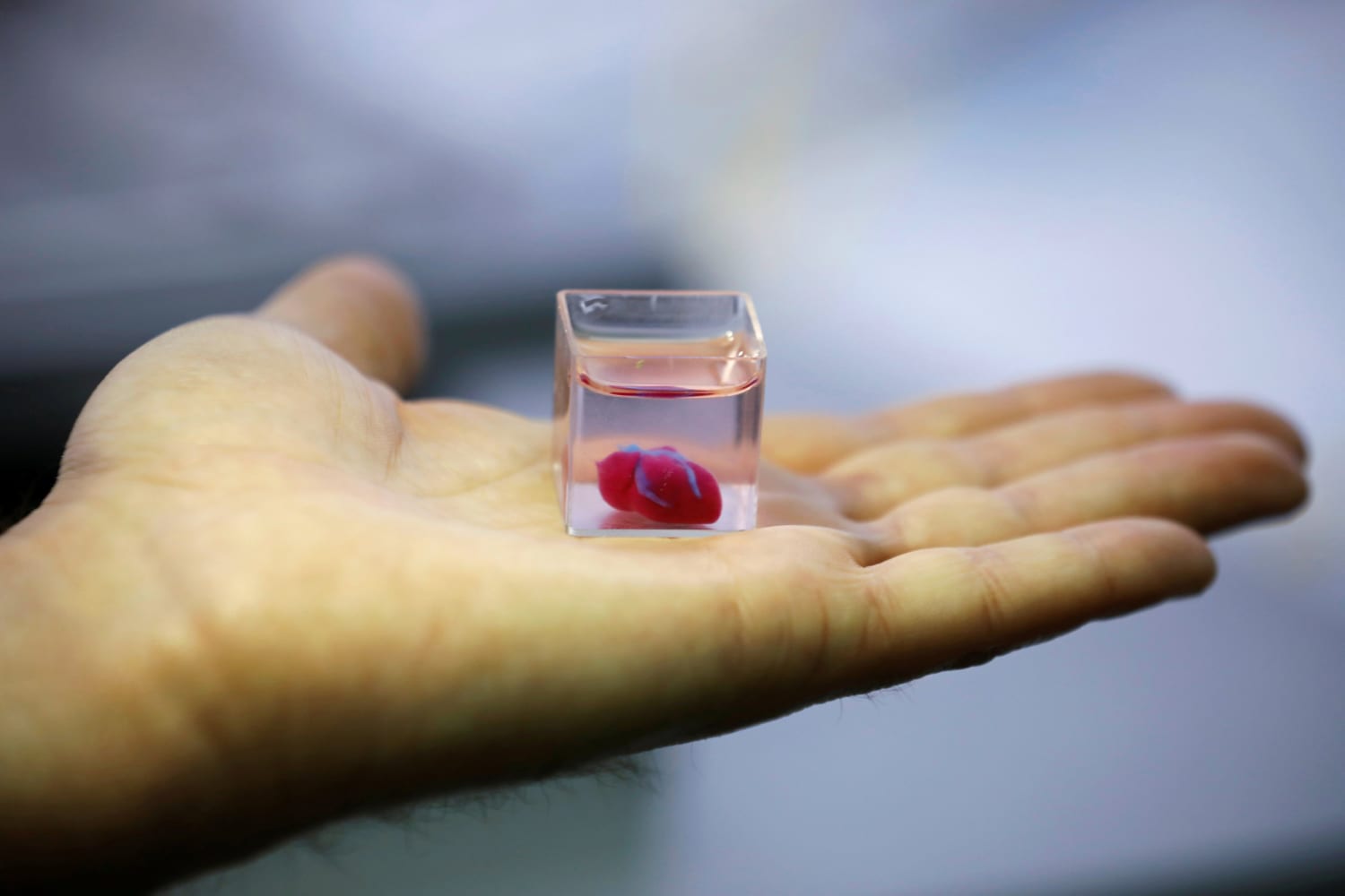
Key Highlights
- Integration of iPSC-derived Cardiomyocytes: Leverage patient-specific and scalable cardiomyocyte production for realistic heart tissue modeling.
- Advanced 3D Bioprinting Techniques: Utilize state-of-the-art bioprinting processes to replicate complex cardiac architectures.
- Enhanced Drug Screening Platform: Develop high-throughput, physiologically relevant models to improve predictive accuracy in pharmacological testing.
Introduction
Traditional drug screening methods that largely rely on 2D cell cultures and animal models often fail to capture the intricacies of human cardiac physiology. This limitation significantly contributes to the inefficiency and safety concerns observed in clinical trials. In response, cutting-edge advancements in 3D printing and bioprinting methodologies have opened new avenues for creating tissue models that closely resemble native human heart structures. This research proposal outlines a comprehensive project to advance the state-of-the-art in 3D printing of human heart models that will transform drug screening procedures by integrating induced pluripotent stem cell (iPSC)-derived cardiomyocytes.
Research Objectives and Overview
The prime objective of this research is to develop and validate a 3D-bioprinted human heart model that exhibits fundamental anatomical and physiological characteristics of native heart tissue. A significant feature of this project is the integration of iPSC-derived cardiomyocytes, which ensures the creation of a physiologically relevant, patient-specific model for enhanced drug efficacy and toxicity screening.
Primary Objectives
- Optimization of iPSC Differentiation Protocols: Develop robust methods for differentiating human iPSCs into mature cardiomyocytes with high purity and consistent function.
- Development and Characterization of Bioinks: Formulate biocompatible bioinks composed of hydrogels such as collagen, gelatin, and hyaluronic acid that support cell viability and mimic the extracellular matrix.
- Establishment of 3D Bioprinting Processes: Optimize printing parameters such as nozzle size, printing speed, and layer resolution to create accurate and structurally stable cardiac tissue models.
- Validation of the Cardiac Model for Drug Screening: Evaluate the 3D-bioprinted cardiac models using a range of pharmaceuticals to assess drug-induced cardiotoxicity and functional responses.
Objective Focus: iPSC-derived Cardiomyocytes
A distinct aspect of our research is to specifically incorporate iPSC-derived cardiomyocytes into the 3D printed heart models. The use of iPSCs allows for:
- Patient-Specific Modeling: Generation of cardiomyocytes from diverse patient sources to simulate personalized cardiac responses.
- Scalability: Reliable, reproducible production of cardiomyocytes in large quantities suitable for high-throughput drug screening.
- Enhanced Physiological Relevance: Creation of cardiac tissue that recapitulates in vivo electrophysiological and contractile properties, essential for accurate drug testing.
Research Design and Methods
1. iPSC Differentiation and Characterization
Procedure
Human induced pluripotent stem cells will be carefully cultured under defined conditions and differentiated into cardiomyocytes using well-established protocols. Differentiation will be monitored and characterized by:
- Flow Cytometry and Immunostaining: Evaluation of cell purity and identification of cardiac-specific markers.
- Electrophysiological Assays: Measurement of action potentials and contractility to ensure functional performance.
2. Bioink Development
Formulation and Testing
The bioink must support both the 3D printing process and the biological requirements of iPSC-derived cardiomyocytes. Development will include:
- Hydrogel Compositions: Testing various combinations of collagen, gelatin, and hyaluronic acid to balance mechanical strength with biocompatibility.
- Printability Evaluations: Optimization based on viscosity and gelation kinetics to ensure high-resolution printing and structural integrity.
3. 3D Bioprinting Process Optimization
Printing Parameters and Architecture
Using advanced bioprinting techniques such as extrusion-based printing and stereolithography, the process will be adjusted to achieve precise control over tissue architecture:
- Printer Modification: Calibration of nozzle diameter, printing speed, and layer thickness aimed at optimizing cell deposition and overall tissue viability.
- Designing Tissue Constructs: Focus on producing models that include accurately aligned cardiac fibers and mimic the coronary vasculature, myocardium, and other structural features.
4. Cardiac Tissue Model and Drug Screening Platform
Model Development
The bioprinted heart models will be cultured under conditions that promote cell maturation and tissue development. Following model creation, the next phase involves:
- High-Throughput Screening: Development of an automated, high-throughput screening platform for rapid drug testing.
- Assay Integration: Utilizing electrophysiological assessments, contractility measurements, and biochemical assays (including cytotoxicity and cardiac injury markers) to assess drug responses.
Data Analysis
Collected data will be analyzed using advanced statistical methods to compare the performance of 3D-bioprinted heart models with traditional 2D cell cultures and animal models. The analysis will address:
- Efficacy: Determining drug-induced improvements or impairments in contractile function.
- Toxicity: Identifying adverse drug effects and establishing dose-response relationships.
Project Timeline and Budget Overview
| Phase | Duration | Key Activities |
|---|---|---|
| Phase 1: Setup and Protocol Development | Month 1-6 | Literature review, iPSC culture establishment, bioink formulation trials. |
| Phase 2: Model Fabrication | Month 7-12 | 3D bioprinting of cardiac models, optimization of printing parameters. |
| Phase 3: Functional Validation | Month 13-18 | Drug screening assays, electrophysiological and contractility measurements. |
| Phase 4: High-throughput Platform Development | Month 19-24 | Integration of automated screening workflows and data analysis pipelines. |
Budget Overview
The estimated total budget for the project is $1.2 million over two years, allocated as follows:
- Personnel: $400,000 (33%)
- Equipment: $300,000 (25%)
- Materials: $200,000 (17%)
- Travel and Training: $100,000 (8%)
- Miscellaneous: $200,000 (17%)
Expected Outcomes and Impact
Anticipated Milestones
- Enhanced Model Realism: A 3D-bioprinted heart model with accurate anatomical and functional traits to serve as an improved platform for drug screening.
- Validated iPSC-derived System: A robust, reproducible protocol for generating patient-specific cardiomyocytes integrated within the heart model.
- Data-Rich Screening Platform: Establishment of a high-throughput screening method that efficiently evaluates drug efficacy and cardiotoxicity.
Broader Impact
This research is expected to significantly enhance the predictive power of preclinical drug testing, reduce the reliance on animal models, and ultimately contribute to safer, more effective cardiovascular therapeutics. The integration of iPSC-derived cardiomyocytes ensures not only improved biological relevance but also opens avenues for personalized medicine approaches, enabling drug development and testing tailored to individual genetic and phenotypic profiles.
References
- Scientific Reports - Nature
- Advances in 3D Bioprinted Cardiac Tissue - Oxford Academic
- Engineered Heart Tissues - The Lancet
-
3D BioPrinting of Cardiac Micro-Tissues - ScienceDirect
-
Functional Tissue Models for Drug Screening - PMC
Recommended
Last updated March 22, 2025