
Research Proposal: Advancing the State-of-the-Art in 3D Printing of Human Heart Models for Enhanced Drug Screening
Innovative biomimetic cardiac tissues for drug screening via advanced bioprinting techniques
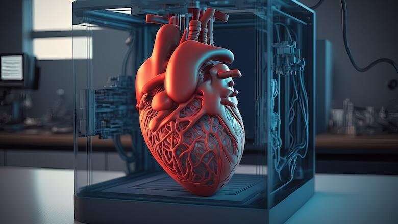
Key Highlights
- Innovative Bioink Formulation: Integration of patient-specific induced pluripotent stem cells (iPSCs) in tailored bioinks for superior cardiac tissue mimicry.
- Multi-Phase 3D Printing Process: Utilization of advanced bioprinting protocols and microfluidic systems that recreate the complex heart microenvironment.
- Enhanced Drug Screening Potential: Development of robust and physiologically accurate models that improve predictability and safety assessments in pharmacological testing.
Introduction and Background
Cardiovascular diseases remain one of the leading causes of mortality worldwide, imposing an enormous burden on healthcare systems. Despite significant advances in therapeutics, the rate of attrition in drug development—mainly due to safety and efficacy issues—demands more reliable preclinical models. Conventional two-dimensional (2D) cell cultures and animal models are frequently inadequate for mimicking the complexities of human heart physiology. As such, advances in three-dimensional (3D) printing technologies have spurred a transformative era in biomedical engineering, with the potential to revolutionize drug screening and personalized medicine.
This research proposal outlines a detailed approach to advance the state-of-the-art in 3D printing of human heart models. By integrating cutting-edge bioprinting methods with sophisticated bioink formulations—including one dedicated to the incorporation of induced pluripotent stem cells (iPSCs)—we propose to develop highly functional in vitro cardiac models. These models aim to not only recapitulate the structural intricacies of human cardiac tissues but also provide dynamic functionalities such as electrical conductivity and mechanical responsiveness. The ultimate objective is to create platforms that allow for enhanced drug screening, reducing dependency on traditional in vivo models and accelerating the drug development process.
Objectives
Primary Goals
The proposed project revolves around several key objectives that collectively aim to elevate the functionality and applicability of 3D printed heart models in drug screening:
1. Development of Advanced Bioink Formulations
The core of this research involves the development of innovative bioinks that effectively mimic the native cardiac extracellular matrix. Central to this is:
- Incorporation of iPSC Cells: A dedicated objective is to optimize the integration of induced pluripotent stem cells (iPSCs) into the bioink. iPSCs will be differentiated into various cardiac cell lineages, including cardiomyocytes, fibroblasts, and endothelial cells, thereby establishing a cell-rich, patient-specific biological milieu. This ensures that the bioink not only provides structural support but also fosters cell survival, differentiation, and functionality.
- Optimization of the mechanical and rheological properties of the bioink to ensure printability while preserving cellular viability and promoting proper tissue organization.
- Tailoring the biochemical cues within the bioink to support interactions among cells, including cell–cell and cell–matrix communication, that are critical for mimicking native cardiac tissue physiology.
2. Fabrication of Functionally Relevant 3D Heart Models
Beyond the bioink formulation, the project emphasizes the development of precise 3D printing protocols that are capable of constructing complex cardiac architectures. This includes:
- Establishing reproducible protocols for layer-by-layer deposition to create models that accurately replicate the 3-dimensional structure of the human heart including chambers, valves, and vascular networks.
- Utilizing advanced bioprinters capable of high-fidelity printing with integrated microfluidic channels to mimic in vivo perfusion and nutrient exchange systems.
- Integrating post-printing maturation processes, such as controlled electrical and mechanical stimulation, to further enhance tissue functionality.
3. Enhanced Evaluation of Drug Responses
A major application of these 3D printed heart models is in the realm of pharmaceutical screening. The research aims to:
- Benchmark the drug responses of 3D printed models against conventional 2D culture systems to demonstrate superior predictive capabilities.
- Implement electrophysiological and mechanical assays to monitor the functional responses of the cardiac models to various drug candidates, ensuring they emulate human pharmacodynamics and pharmacokinetics accurately.
- Create high-throughput screening protocols that can facilitate rapid testing of multiple compounds, accelerating the preclinical evaluation process.
4. Integration of Microfluidics and Biomaterials for a Realistic Cardiac Microenvironment
It is essential that the in vitro models recreate the dynamic microenvironment of the human heart. To this end, the proposal incorporates:
- Designing microfluidic systems that deliver precise gradients of nutrients, oxygen, and mechanical stimuli to the printed tissues.
- Utilizing biomaterials with tunable properties that simulate the native stiffness and elasticity of myocardial tissue, thereby promoting realistic cell behavior.
- Incorporating vascular structures within the printed constructs to ensure adequate perfusion and mimic the circulatory dynamics of native heart tissue.
5. Validation through Multimodal Functional Assessments
To ensure the developed heart models have robust applications in drug screening, they will undergo comprehensive validation:
- Utilizing optical mapping and electrophysiology to confirm the presence of coordinated electrical signals similar to those observed in natural cardiac tissues.
- Conducting biomechanical tests to evaluate tissue contractility, strength, and resilience under physiological stresses.
- Applying advanced imaging techniques to monitor cellular organization, tissue integration, and long-term viability during drug exposure experiments.
Methodology
Step-by-Step Process
Phase 1: iPSC Isolation, Differentiation, and Bioink Preparation
The first phase involves isolating iPSCs from patient-specific sources followed by differentiation into key cardiac cell types. These cells will then be incorporated into a customized bioink formulation. This phase includes:
- Collection and reprogramming of patient-derived somatic cells to generate iPSCs.
- Application of specific growth factors and small molecules to induce the differentiation of iPSCs into cardiomyocytes, endothelial cells, and cardiac fibroblasts.
- Formulation of a bioink that combines these differentiated cells with hydrogels such as alginate, gelatin methacrylate (GelMA), or other biocompatible materials optimized to mimic the extracellular matrix of cardiac tissue.
- Characterization of the bioink for viscosity, cross-linking potential, and cell viability to ensure it meets the technical demands of the bioprinting process.
Phase 2: 3D Bioprinting of Cardiac Constructs
This phase focuses on the production of the heart models utilizing a state-of-the-art 3D bioprinter:
- Development of CAD models that define the intricate architecture of human heart structures such as the atria, ventricles, valves, and embedded vasculature.
- Optimization of printing parameters (e.g., nozzle diameter, printing speed, and layer thickness) to ensure high-fidelity reproduction of the designed structures.
- Integration of microfluidic elements and spatial patterning that allow for tissue perfusion and incorporate realistic gradients of oxygen and nutrients.
- Post-printing processes that include controlled maturation environments providing electrical and mechanical stimuli to promote tissue maturation and functional integration.
Phase 3: Functional Validation and Drug Screening
The final phase encompasses extensive functional assessments and the evaluation of the models’ responsiveness to pharmacological agents:
- Employing electrophysiological assays to measure action potentials and conduction velocities which are indicative of healthy cardiac function.
- Performing contractility tests using optical mapping and imaging methodologies to assess the mechanical performance of the tissues under simulated physiological conditions.
- Running a series of drug screening experiments where the tissue models are exposed to different cardiovascular agents, with subsequent analysis of cellular responses, electrophysiological alterations, and overall tissue viability.
- Comparative analyses between 3D printed tissues and conventional 2D cultures to validate the advantages in predictive pharmacodynamics and toxicity assessments.
Comprehensive Project Timeline and Resource Allocation
A clear timeline alongside dedicated resource allocation is imperative for successful project execution. The following table outlines a detailed schedule covering all phases of the research:
| Phase | Duration | Key Activities |
|---|---|---|
| Phase 1 | Months 1-3 | iPSC isolation and differentiation, bioink development, rheological testing |
| Phase 2 | Months 4-6 | 3D bioprinter parameter optimization, fabrication of cardiac constructs, integration of microfluidics |
| Phase 3 | Months 7-9 | Functional assays, electrophysiological mapping, drug response evaluation |
| Phase 4 | Months 10-12 | Data analysis, refinement of protocols, manuscript preparation, and validation |
Resource Requirements and Budget Estimates
The project requires substantial investment in both hardware and human resources. Estimated budgetary allocations include:
- Equipment: Acquisition of advanced 3D bioprinters, microfluidic devices, and bio-reactors.
- Materials and Reagents: Procurement of high-quality biocompatible materials, hydrogels, growth factors, and iPSC culture reagents.
- Personnel Costs: Funding for specialized researchers, postdoctoral fellows, and technical support to conduct experimentation and data analysis.
- Miscellaneous: Allocations for consumables, maintenance, and collaboration with pharmaceutical partners for validation studies.
Potential Impact and Future Applications
Innovative Transformations in Drug Screening
The successful realization of this proposal is poised to transform both the field of cardiac tissue engineering and drug development. By using iPSC-laden bioinks and advanced 3D printing protocols, the resulting models offer unparalleled accuracy in mimicking the human heart. This, in turn, enhances the reliability of preclinical testing, allowing for:
- Early detection of adverse drug reactions and cardiotoxicity, mitigating risks before clinical trials.
- Personalized drug screening where patients’ own cells guide the development of tailored therapeutic strategies.
- Accelerated discovery and approval pathways for new cardiovascular drugs, ultimately reducing the time-to-market for critical treatments.
Furthermore, the methodologies and technological advancements conceived through this research can be extrapolated to other tissues and organs, fostering a broader spectrum of applications in regenerative medicine and personalized healthcare.
References
-
3D Bioprinting for Cardiovascular Tissue Engineering - PMC
-
Advanced Bioink Formulations with iPSCs - NIH PMC
-
Realistic Cardiac Tissue Models for Drug Testing - ScienceDirect
- 3D Bioprinting at the Frontier of Regenerative Medicine - Frontiers
- 3D Bioprinting Platform for Cardiac Tissue Models - AIP
Recommended Further Queries
Last updated March 22, 2025